Men have always struggled with initiating, manipulating and maintaining erections. The erections are necessary for several years for sexual, pleasure and reproductive functions.
Problems with erection may indicate that either youre not healthy or your bad erections will someday eat you up.
Here is the list of vitamins that are found to be useful by different researches done at several reputed universities and research centers worldwide.
Helps men with weak erection by; – Improving blood flow – Reducing inflammation – Supporting testosterone levels.
Here’s a list of foods that are good sources of vitamin B3 (niacin):
Animal Sources:
Meat:
Chicken (especially chicken breast)
Turkey
Beef (especially liver)
Fish:
Tuna
Salmon
Mackerel
Dairy Products:
Milk
Yogurt
Cheese
Plant Sources:
Legumes:
Peanuts
Lentils
Beans (black beans, kidney beans)
Nuts and Seeds:
Sunflower seeds
Almonds
Walnuts
Whole Grains:
Brown rice
Oats
Barley
Whole wheat bread
Vegetables:
Mushrooms (especially portobello and shiitake)
Potatoes
Green peas
Fruits:
Avocado
Bananas
Fortified Foods:
Breakfast cereals
Nutritional yeast
Including a mix of these foods can help you meet your vitamin B3 needs!
2. Vitamin D
Helps men by:
Regulating blood pressure –
Improving blood flow to the penis –
Supporting hormone regulation.
This can lead to improved erectile function and overall sexual health.
3. Vitamin C 100mg
Helps men with weak erections by; – Boosting nitric oxide – Reducing oxidative stress – Improving blood flow This leads to improved erectile function.
Here’s a comprehensive list of foods that are excellent sources of vitamin C:
Fruits:
Citrus Fruits:
Oranges
Grapefruits
Lemons
Limes
Berries:
Strawberries
Raspberries
Blueberries
Blackberries
Other Fruits:
Kiwi
Pineapple
Mango
Papaya
Watermelon
Cantaloupe
Guava
Acerola cherries
Vegetables:
Peppers:
Bell peppers (especially red and yellow)
Chili peppers
Leafy Greens:
Kale
Spinach
Swiss chard
Mustard greens
Cruciferous Vegetables:
Broccoli
Brussels sprouts
Cauliflower
Root Vegetables:
Sweet potatoes
Carrots
Tomatoes:
Fresh tomatoes
Tomato juice
Tomato sauce
Herbs:
Fresh parsley
Cilantro
Thyme
Other Sources:
Fortified juices (like orange juice)
Fortified foods (some breakfast cereals)
Incorporating a variety of these fruits and vegetables into your diet can help you meet your vitamin C needs effectively!
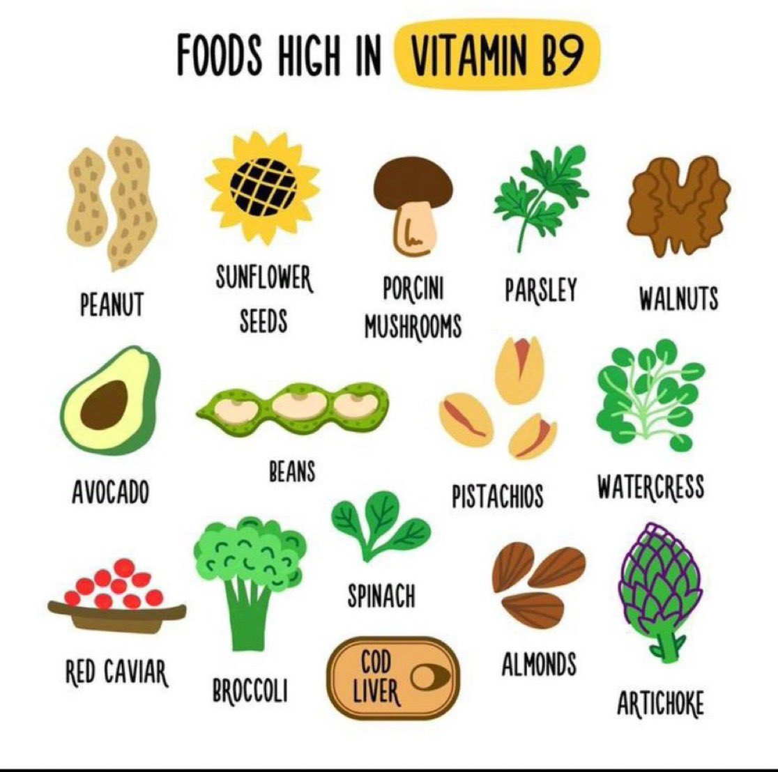
4. Vitamin B9 ( Folic Acid)
Helps men with weak erections by; – Reducing homocysteine – Supporting nitric oxide production This helps to improve erectile function.
Here’s a list of foods that are good sources of vitamin B9 (folate):
Leafy Greens:
Spinach
Kale
Swiss chard
Legumes:
Lentils
Chickpeas
Black beans
Fruits:
Oranges
Bananas
Avocado
Nuts and Seeds:
Sunflower seeds
Peanuts
Almonds
Whole Grains:
Quinoa
Brown rice
Fortified cereals
Vegetables:
Broccoli
Brussels sprouts
Beets
Animal Products:
Liver (beef or chicken)
Eggs
Fortified Foods:
Bread
Pasta
Breakfast cereals
Including a variety of these foods in your diet can help ensure you get enough vitamin B9!
5. Vitamin B12
Helps men with weak erection by; – Supporting nerve function – Reducing homocysteine.
If you’re overlooking B12, you’re holding yourself back. Proper nerve function and healthy blood flow aren’t just ‘nice to haves’ They’re essentials for strength in every area.
6. Vitamin L- Carnitine
It helps men with weak erection by – Boosting energy and sperm health – Supporting nitric oxide production
Here’s a list of foods that are good sources of L-carnitine:
Meat:
Beef (especially red meat)
Lamb
Pork
Poultry:
Chicken
Turkey
Fish:
Cod
Tuna
Salmon
Dairy Products:
Milk
Cheese
Yogurt
Vegetables (in smaller amounts):
Avocado
Asparagus
Spinach
Whole Grains:
Whole wheat bread
Oats
While animal products are the richest sources of L-carnitine, small amounts can also be found in some plant-based foods.
7. Zinc
It helps men with weK erections by – Enhancing sperm quality – Boost immune function – Supporting testosterone production
Here’s a list of foods that are good sources of zinc:
Meat:
Beef
Pork
Lamb
Poultry:
Chicken
Turkey
Seafood:
Oysters (one of the highest sources)
Crab
Lobster
Shrimp
Dairy Products:
Milk
Cheese
Yogurt
Legumes:
Chickpeas
Lentils
Beans
Nuts and Seeds:
Pumpkin seeds
Cashews
Hemp seeds
Whole Grains:
Quinoa
Brown rice
Oats
Fortified Foods:
Breakfast cereals
Snack bars
Vegetables (in smaller amounts):
Spinach
Mushrooms
Broccoli
Including a variety of these foods in your diet can help ensure adequate zinc intake!
8. Vitamin B6
Helps men with weak erection by – Regulating hormones – Supporting neurotransmitters – Reducing stress and anxiety
Meat:
Chicken (especially chicken breast)
Turkey
Beef liver
Fish:
Salmon
Tuna
Halibut
Pulses:
Chickpeas
Lentils
Black beans
Nuts and Seeds:
Sunflower seeds
Pistachios
Hazelnuts
Whole Grains:
Brown rice
Oats
Barley
Fruits:
Bananas
Avocado
Watermelon
Vegetables:
Potatoes
Spinach
Sweet potatoes
Fortified Foods:
Breakfast cereals
Nutritional yeast
Incorporating a mix of these foods can help you meet your vitamin B6 needs!
If you have been experiencing erectile disfunction then that is the way you can solve it. Very simple and clear don’t struggle again.
कार्डियोलोजीमा, सिपि आर (CPR) भनेको “कार्डियोपल्मोनरी रेससिटेशन” हो। यो एक आपतकालीन प्रक्रिया हो जुन कसैको हृदय र श्वासप्रश्वास अचानक बन्द भएमा प्रयोग गरिन्छ। CPR ले छातीमा थिचेर र कृत्रिम श्वास दिँदै गर्दा रगत र अक्सिजनको प्रवाहलाई कायम राख्न मद्दत गर्छ, जसले गर्दा मस्तिष्क र अन्य महत्वपूर्ण अंगहरूसम्म अक्सिजन पुग्न सक्छ।
meaning of cpr in nepali
कार्डियोपल्मोनरी रेससिटेशन एक आपतकालीन प्रक्रिया हो जुन कसैको हृदय र श्वासप्रश्वास अचानक बन्द भएमा छातिमा थिचेर र कृत्रिम श्वास दिँदै गर्दा रगत र अक्सिजनको प्रवाहलाई कायम राख्न मद्दत गर्छ।
Here in this video we will explain how to do CPR in Nepali.
Performing CPR in a dummy
CPR गर्दा निम्न चरणहरू अपनाइन्छ:
सुरक्षितता सुनिश्चित गर्नुहोस्: घटना स्थल सुरक्षित छ कि छैन जाँच गर्नुहोस्।
प्रतिक्रिया जाँच गर्नुहोस्: पीडितलाई हल्का टोक्नुहोस् र बोलाउनुहोस्।
एम्बुलेन्स बोलाउनुहोस्: यदि पीडितले कुनै प्रतिक्रिया दिँदैन भने तुरुन्तै आपतकालीन सेवामा सम्पर्क गर्नुहोस्।
छाती थिच्ने: पीडितको छातीको बीचमा आफ्नो हात राखेर तीव्र र गहिरो थिच्ने गर्नुहोस् (प्रति मिनेट 100-120 पटक)।
कृत्रिम श्वास: यदि सम्भव छ भने, हावा दिने माध्यमबाट प्रत्येक 30 थिचाईपछि दुई पटक हावा फुकेर श्वास दिनुहोस्।
CPR को सही ज्ञान र अभ्यासले धेरै व्यक्तिहरूको जीवन बचाउन सक्ने क्षमता राख्दछ।
सिपि आर गर्ने तरिका भिडियो How to do CPR in Nepali
राम्रो सिपिआर (CPR) का फाइदाहरू मुटुको स्वास्थ्यको सन्दर्भमा निम्न प्रकारका छन्:
जीवन बचाउने क्षमता:
जब कसैको मुटु रोक्छ (कार्डियक अरेस्ट), तुरुन्तै सिपिआर दिनाले रगत र अक्सिजनको प्रवाहलाई कायम राख्न मद्दत गर्छ, जसले गर्दा मस्तिष्क र अन्य महत्वपूर्ण अंगहरूमा अक्सिजनको अभाव हुनबाट बचाउँछ।
समयको महत्वपूर्ण भूमिका:
सिपिआर तुरुन्त सुरु गर्नु महत्त्वपूर्ण छ। मुटु रोकिएको ३-५ मिनेटभित्र सिपिआर दिनाले बच्ने सम्भावना धेरै बढाउँछ।
मस्तिष्कको क्षति कम गर्नु:
मस्तिष्कलाई अक्सिजनको आवश्यकता हुन्छ। सिपिआरले मस्तिष्कमा अक्सिजनको आपूर्ति कायम राख्न मद्दत गर्छ, जसले गर्दा स्थायी मस्तिष्क क्षति कम गर्न सक्छ।
पुनः मुटुको चाल फिर्ता ल्याउन सहयोग:
सिपिआरले मुटुको चाललाई पुनः सुरु गर्न मद्दत पुर्याउँछ र पछि एइडी (AED) जस्ता उपकरणहरूको प्रयोगले थप सहयोग पुर्याउन सक्छ।
हस्पिटल पुग्न समय दिनु:
सिपिआरले बिरामीलाई अस्पतालमा पुग्न समय दिन्छ, जहाँ विशेषज्ञहरूले थप उपचार दिन सक्छन्।
रक्तसञ्चारलाई कायम राख्नु:
सिपिआरले शरीरमा रक्तसञ्चारलाई कायम राखेर विभिन्न अंगहरूमा अक्सिजन र पोषक तत्वहरूको प्रवाहलाई निरन्तरता दिन्छ।
समग्रमा, सिपिआर एक अत्यावश्यक जीवन रक्षक प्रक्रिया हो, जसले मुटुको आकस्मिक रोकावट हुँदा मान्छेको जीवन बचाउन महत्वपूर्ण भूमिका खेल्छ। सिपिआरको सही तालीम लिनु र सिपिआरको प्रक्रिया राम्रोसँग जान्नु सबैका लागि आवश्यक छ।
Title: How to Perform a Good CPR in Nepali (Video 2081): CPR Technique in Nepali
राम्रो सिपिआर गर्ने तरिका (नेपालीमा)
सुरक्षित स्थान सुनिश्चित गर्नुहोस्:
सर्वप्रथम, तपाईँले जहाँ सिपिआर गर्न लाग्नु भएको छ, त्यो स्थान सुरक्षित छ कि छैन जाँच गर्नुहोस्। दुर्घटनाको सम्भावना भएमा, बिरामीलाई सुरक्षित स्थानमा सार्नुहोस्।
चेतना जाँच गर्नुहोस्:
बिरामीलाई काँधमा हल्का हल्लाएर र सोधेर “के तपाईँ ठीक हुनुहुन्छ?” जस्ता प्रश्न सोध्नुहोस्। कुनै प्रतिक्रिया नभएमा तुरुन्तै आपतकालीन नम्बरमा फोन गर्नुहोस्।
श्वासप्रश्वास जाँच गर्नुहोस्:
बिरामीको नाक र मुख नजिक कान राखेर श्वासप्रश्वास जाँच गर्नुहोस्। श्वासप्रश्वास नभएमा वा असामान्य भएमा, सिपिआर सुरु गर्नुहोस्।
छातीको कम्प्रेसन (Compressions) गर्नुहोस्:
बिरामीलाई सपारेर सिधा सम्याउनुहोस्।
तपाईँको एक हातको हत्केलालाई बिरामीको छातीको बिच भागमा राख्नुहोस् र अर्को हातलाई माथि राखेर अड्याउनुहोस्।
३० पटक छातीलाई ५-६ सेमी तलसम्म थिच्नुहोस्। प्रति मिनेट १००-१२० पटकको गतिमा गर्नुहोस्।
श्वासप्रश्वास दिनुहोस् (Ventilations):
बिरामीको टाउकोलाई पछाडि झुकाएर, च्यापिएको नाकलाई बन्द गरेर मुखबाट दुई पटक श्वास दिनुहोस्।
हरेक श्वास १ सेकेन्डसम्म दिनुहोस् र छाती उठेको देख्नुभयो भने मात्र अर्को श्वास दिनुहोस्।
क्रम दोहोर्याउनुहोस्:
३० पटक छाती थिचेपछि २ पटक श्वास दिने क्रमलाई निरन्तरता दिनुहोस्।
जबसम्म बिरामीले श्वास लिन सुरु गर्दैन वा आपतकालीन सहायता आइपुग्दैन, सिपिआर निरन्तर गर्नुहोस्।
सिपिआर गर्दा ध्यान दिनुपर्ने महत्वपूर्ण कुराहरू:
छातीको कम्प्रेसन गहिरो र द्रुत गर्नुपर्छ।
श्वासप्रश्वास दिनुअघि छाती थिचेको सुनिश्चित गर्नुहोस्।
प्रक्रिया गर्दा थकित भएमा, अर्को व्यक्ति तयार राख्नुहोस्।
Snake bite piercings have been made since lomg ago but they had never been into medical attention till lately when there had been cases of multiple medical illnesses and side effects with extreme cases of snake bite piercings.
Snake bite piercing are named after what they look like that is Fangs of a snake, or the bitemark of a snake.
The side effects and complications of snake bite piercings are listed down below in different heading.
Hence while they look pretty for what they are they can be devastatingly ugly like what they are named after.
There are typically Three location where the snake bite piercings can be kept. They include tongue, midline on lower lip, one side on lower lip and both side on lower lip.
A strict sterile precaution should be followed while making a snakebite piercing. This includes proper sterility of the sharps, wearing sterile gloves and sterile piercing site preparation.
The procedure starts by preparing all these instruments and then a sterile field is prepared. With sterile precaution the piercing is done and a sterile temporary assessory is put on in the piercing.
Additionally, an antiseptic/antibiotic preparation might be applied post procedure. These products include Mupirocin, Polysporin, Betadine, Chlorhexidine etc.
No dressing i applied and the wound is left open to heal. If there are any signs of infection seen like pain, swelling, redness, discharge etc you should contact your doctor immidiately.
The person doing this prcedure should be extra cautious not to prick htemselves and should make sure that the client does not have any infections like HIV, Hepatitis (B and C), syphillis etc.
Managing Pain and Discomfort: Medical Insights for Snake Bite Piercing
The pain management can be achieved via application of local anesthetic. Local anesthetic of choice is lignocaine with or without adrenaline (1-2%).
The client can be adviced to take over the counter (OTC) pain medication like Ibuprofen, Paracetamol (Acetaminophen), naproxen, ketorolac, diclofenac or aceclofenac (prescription).
Healing and Aftercare: Medical Recommendations for Snake Bite Piercings
It’s important to note that while there are plenty of risks, proper care, hygiene, and following the guidance of a professional piercer can help minimize these potential risks and complications.
Remember that, it’s important to consult with a healthcare professional or a qualified well trained piercer for personalized advice and guidance made for your specific situation and need.
The healing of the piercing can sometimes take longer than told by the piercer. But you shouldnt panic and wait for a couple or more days. If it still doesn’t heal then you should immidiately seek advice from a healthcare professional.
Potential Risks and Complications of Snake Bite Piercings: A Medical Perspective
Infection
Like any other piercing, snake bite piercings also carry a risk of infection. Improper aftercare, unsterilized equipment, or poor hygiene can increase the risk of infection.
Swelling and Bruising
Initial swelling and bruising are common after getting snake bite piercings. Excessive swelling can potentially lead to long term discomfort and difficulties with eating or speaking.
Rejection or Migration
There is a possibility that the body may reject the snake bite piercings, leading to the piercing4 being pushed out of the skin or the piercing shifting from its original position.
Gum and Tooth Damage
Snake bite piercings located near the lower lip can come into contact with the gums and teeth. Continuous contact or improper jewelry size can result in gum recession, enamel erosion, chipped teeth, or damage to dental work.
Allergic Reactions
Some individuals may have allergic reactions to certain metals used in snake bite piercing jewelry, particularly if they have sensitivities or allergies to nickel or other common metals. Allergic reactions can manifest as redness, itching, swelling, or irritation around the piercing site. Long-Term Effects and Oral Health Considerations of Snake Bite Piercings
Snake bite piercing tongue is one of the common tngue piercing. This has been done by many famous celibrities aroud the world and has successfully been on the market for long time.
2. Viper bites piercing
Viper bites piercing is a type of snake bite piercing named after a famous poisonous snake Viper. This signifies this piercing is as furious as viper.
3. Multiple Snakebite Piercing
Multiple snake bite piercing is one type of piercing commonly done in a goth culture. There can be more than one in lower lip.
4. Midline Snakebite piercing
Piercing in the midline lip is most beautiful and is done by somany more celebrities as well.
full ring or half ring piercing with diamond are also equally famous.
5. Single unilateral Snakebite piercing/one side Snake bite piercings
Unilateral piercing is beautiful like damn. The side look or even the front look is so pretty and will make you attractive.
This doen’t make you uncomfortble and doesn’t affect you speech either.
FAQs
Are snake bite piercings painful?
Generally no. But Snakebite piercings can occasionally be painful if done inappropriately, infected or pain management or anesthesia is not done properly.
How long does it take for snake bite piercings to heal?
It usually taked 5 to 10 days. Contact your doctor if it is taking longer.
What are the potential risks and complications associated with snake bite piercings?
Potential risks include pain, bleeding, infection, allergy, rejection and disfigurement. Discuss with your piercer prior to procedure.
Can snake bite piercings damage teeth or gums?
Piercing itself may not damage it. But of you use low quaity and allergen, chemical containing piercings your teeth and gums may damange. Occasionally the damage ay occur by using unnecessarily large, uncomfortable and bad quality built ornament.
Are there any specific aftercare instructions for snake bite piercings to prevent infection?
Strict infection prevention and sterile precaution should be followed by the piercer and the client.
AHAs and BHAs are the types of hydroxy acids that help in exfoliating the skin. AHA and BHA are type of medical skincare products that are found in multiple skin care products like moisurizers, creams, scrubs, peelers, cleansers, toners, masks etc.
AHA fullform: Alpha hydroxy acid
BHA full form: Beta hydroxy acids
PHA full form: Polyhydroxy acids
They are hydroxy acids that are derived from fruit products.
After use of AHA BHA PHA skincare products you’ll gradually start feeling that your skin is softer than before.
What is AHA BHA toner
AHA BHA PHA toners are the types of toners that help in exfoliating the outermost layer of skin and in the hair follicles and sebaceous glands.
This helps in clearing the dead tissue and help the skin get proper exposure to outer environment and helps make skin look better.
AHA vs BHA skincare products
Read more regarding AHA vs BHA in this healthline website link.
There is no exact difference between AHA BHA and PHA composition products and theres none better than others. The common differences can be seen below.
In general, AHA has anti aging effects, anti drying effets and BHA has tone maintainance property.
Paula’s choice aha bha toner (Paula’s choice toner)
Paula’s choice has been one of the long standing and doctor’s recommended skin peeler available in the market. Toda it ias been one of the top choices due to its good quality and effective results. There are minimal side effects with proper use and is relatively cost-effective as well.
Can i use cosrx aha bha toner with vitamin c? Answer: Yes you can. But try to alternate the time when you apply the product. Apply one at nightime while other at morning or vice versa.
Can i use cosrx aha bha toner everyday? Yes you can. But make sure youre not getting side effects and your skin has not thinned a lot. If youthink youre using it a lot then contact your dermatologist. Yoou can contact doctor at our site or here.
What are the side effects of skin peelers? They may irritate your skin, make it dry and may cause photosensitivity. Please consult your doctor regarding this.
What amount should I apply? The dose is not fixed but individual. You should apply the amunt that makes very thin layer in your face and is not excess. Always protect your eyes from the product as these products may harm your eyes.
What should I do if I am pregnant? These skincare products are NOT recommended for pregnant women if they contain retinol (vitamin A1). The products containing AHA, BHA and PHA are generally safe for pregnant woman. Please consult your doctor if you’re pregnant.
When it comes to our health, paying attention to even the smallest symptoms is vital. Chest pain is often associated with heart problems, however did you recognize that arm pain can also be a potential indicator? In this article, we are able to discover the relationship between arm pain and heart troubles, supporting you recognize while to are trying to find medical attention and a way to stay proactive approximately your cardiovascular health.
Chest pain with heart disease
Understanding Arm pain
Arm pain can be one of the symptoms of heart disease. Arm ache/pain can be experienced in various methods, along with aching, pulsating, or a pins-and-needles sensation. Even as no longer all times of arm ache indicate coronary heart troubles, it is crucial to recognize while it may probably be a symptom of an underlying cardiovascular issue.
Possible causes of pain to the left arm
One of the many causes of left arm pain is heart disease but it is not so common. More then that isolated left sided arm pain can be less likely due to the heart disease rather it can be musculoskeletal or neurological cause. So, it’s vital to notice that arm pain can also be resulting from factors like muscle pressure, injury, or stress on nerves. For an accurate diagnosis, it’s recommended to consult with a healthcare expert.
Pain and coronary heart diseases
Arm pain as a Symptom of coronary heart issues it’s important to note that not all people experiencing coronary heart problems will have arm ache, and now not all arm ache indicates a heart condition. however, arm ache, especially in the left arm, is typically related to coronary heart issues. that is because the nerves from the coronary heart and the arm share pathways inside the spinal twine, that may lead to referred ache.
Left sided chest pain with heart disease
Nature of pain in heart disease
Types of heart situations and Arm ache a) heart assault: throughout a heart attack, blood drift to the heart is blocked, inflicting chest ache or pain. This pain can radiate to the left arm, shoulder, or even the jaw. if you revel in excessive, chronic, or unexpected arm pain in conjunction with other heart assault symptoms like chest pain, shortness of breath, and dizziness, are trying to find instant medical attention.
The pain of heart attack usually last more than 15 minutes and so arm pain of heart attack also lasts more than 15 minutes. To know your arm pain is heart related you should have other risk factors as well as heart disease symptoms.
What does heart attack arm pain feel like?
Heart attack pain is usually continuous type, vague and pressing type. It will be associated with central chest, left chest, jaw, neck , or upper abdominal pain, It is also associated with shortness of breath, weakness and sweating. Usually the pain is very severe.
Is always left arm involved in heart attack?
No. It is mostly left arm but sometimes the pain might be radiated to the right arm as well during the heart attack and shoul we be equaly cautious regarding to the right arm as well. Ignoring this fact even the highly talented healthcare personnel may miss the diagnosis leading to bad outcomes.
Arm pain and heart attack: which arm hurts during heart attack?
When to are trying to find scientific interest arm pain on my own might not continually suggest a coronary heart trouble, however in case you enjoy any of the subsequent signs similarly to arm ache, it is important to seek on the spot scientific interest:
6 Symptoms of heart disease
Chest pain or discomfort
Shortness of breath
Sweating
Nausea or vomiting
Lightheadedness or fainting
Pain that radiates down the left arm, jaw, or shoulder
At the same time as arm pain may have various causes, it is essential to be aware about the capacity connection among arm ache and coronary heart problems. understanding the signs and symptoms associated with heart conditions, including chest pain and arm ache, permit you to make informed choices about in search of scientific assistance. remember, it is always higher to misunderstand the side of caution and seek advice from a healthcare expert when you have issues about your heart fitness.
People with red hair need more anesthesia: Research review
Do redheads need more anesthesia than others?
What does the biology and medical science say?
Recently I was going through an online social network and I saw a post saying redheads need more aneshthesia and here’s review on the topic based on multiple researches and publications.
Hope youll enjoy reading it.
The Science Behind Why Redheads Need More Anesthesia
Have you ever wondered why redheads may require more anesthesia than people with other hair colors? It turns out that there is a scientific explanation for this phenomenon. Redheads have a genetic variation that affects the way their bodies process pain and anesthesia.
The mutation in melanocortin-1 receptor has been attributed to the red hair of theose people and same for the difference in the sensivity to the pain and depth of anesthesia as well.
This variation causes their bodies to be more resistant to pain medication, requiring higher doses to achieve the same level of pain relief. In addition, redheads also have a higher likelihood of experiencing anxiety and nervousness before surgery, which can also affect their response to anesthesia. This unique attribute of redheads has been studied extensively by medical professionals, and understanding this genetic variation can help anesthesiologists provide more effective care for their redheaded patients. So, if you’re a redhead or know someone who is, read on to discover the fascinating science behind this phenomenon.
What is anesthesia and how does it work?
There are three types of anesthesia: general, regional, and local. The anesthesia described here may refer to local or regional anesthesia, sedation and general anesthesia.
General anesthesia is a state of induced unconsciousness that is used to provide pain relief and muscle relaxation during medical procedures. General anesthesia is the most common and involves the use of drugs that put the patient to sleep and block pain signals to the brain. Regional anesthesia involves numbing a specific area of the body, while local anesthesia involves numbing a small area, such as a tooth or skin patch.
The drugs used in anesthesia work by blocking the transmission of nerve signals that communicate pain and other sensations to the brain. This allows the patient to undergo medical procedures without feeling any pain or discomfort. The mechanism of general enesthesia like propofol may not be completely understood r explained. The amount of anesthesia required depends on various factors, including the patient’s weight, age, and medical history.
The genetic mutation responsible for red hair
Red hair is caused by a genetic mutation in the MC1R gene, which is responsible for producing the pigment that gives hair its color. This mutation causes the gene to produce a protein that is less effective at producing the pigment, resulting in red hair. The MC1R gene is also responsible for producing melanin, which is the pigment that gives skin its color. People with the red hair gene mutation have less melanin in their skin, which makes them more susceptible to sunburn and skin cancer.
The MC1R gene mutation is inherited in an autosomal recessive pattern, meaning that a person must inherit two copies of the mutated gene (one from each parent) to have red hair. However, even people with just one copy of the mutated gene may exhibit some of the traits associated with red hair, such as fair skin and freckles.
The link between red hair and anesthesia: Does being a redhead affect anesthesia?
Research has shown that people with red hair require more anesthesia than people with other hair colors. This is because the MC1R gene mutation affects the way the body responds to certain drugs, including pain medication and anesthesia. The mutation causes the body to produce less of a protein called POMC, which is involved in the production of endorphins, the body’s natural painkillers. This results in redheads being less sensitive to certain types of pain medication and requiring higher doses to achieve the same level of pain relief.
In addition to being less sensitive to pain medication, redheads also have a higher likelihood of experiencing anxiety and nervousness before surgery. This can be attributed to the fact that the same genetic mutation that causes red hair also affects the production of the stress hormone cortisol. People with the mutation produce less cortisol, which can lead to increased anxiety and stress.
Studies on redheads and anesthesia: Does anesthesia affect hair color?
Several studies have been conducted to investigate the link between red hair and anesthesia. One study published in the British Journal of Anaesthesia found that redheads required 20% more anesthesia than people with other hair colors. Another study published in the journal Anesthesiology found that redheads required 19% more anesthesia than people with dark hair and 16% more than people with blonde hair.
These studies also found that redheads were more likely to experience side effects from anesthesia, such as nausea and vomiting. This may be due to the fact that the drugs used in anesthesia are metabolized differently in people with the MC1R gene mutation, leading to a longer recovery time and increased risk of side effects.
Redhead people require more anesthesia: Anesthesist providing anesthesia
Differences in anesthesia dosage for redheads
The fact that redheads require more anesthesia than people with other hair colors has important implications for healthcare providers. Anesthesiologists must take this into account when administering anesthesia to redheaded patients, as failing to do so can result in inadequate pain relief and increased risk of side effects.
To ensure that redheads receive the appropriate amount of anesthesia, anesthesiologists may need to adjust the dosage based on the patient’s hair color and genetic makeup. This can be done by conducting a genetic test to determine whether the patient has the MC1R gene mutation and adjusting the anesthesia dosage accordingly.
Other medical considerations for redheads
In addition to requiring more anesthesia, redheads may also be more susceptible to certain medical conditions. For example, research has shown that redheads have a higher risk of developing skin cancer due to their fair skin and reduced ability to produce melanin. They may also be more susceptible to certain types of pain, such as chronic pain and migraine headaches.
Redheads may also require different dosages of other types of medication, such as painkillers and antidepressants. This is because the MC1R gene mutation affects the way the body processes these drugs, just as it affects the way the body processes anesthesia.
Challenges for healthcare providers
The fact that redheads require more anesthesia and may be more susceptible to certain medical conditions can present challenges for healthcare providers. Anesthesiologists must be aware of the unique needs of redheaded patients and take steps to ensure that they receive the appropriate care.
This may involve conducting genetic testing to determine whether the patient has the MC1R gene mutation and adjusting the anesthesia dosage accordingly. It may also involve providing additional support to redheaded patients who may be more anxious or stressed before surgery.
Coping mechanisms for redheads undergoing anesthesia: incomplete anesthesia
If you’re a redhead who is scheduled to undergo anesthesia, there are steps you can take to help ensure a successful outcome. First, make sure to inform your healthcare provider that you have red hair and discuss any concerns you may have about anesthesia. Your provider can work with you to develop a plan that addresses your unique needs and concerns.
You may also want to consider relaxation techniques such as deep breathing, meditation, or visualization to help reduce anxiety and stress before surgery. These techniques can help you feel more relaxed and may even reduce the amount of anesthesia you require.
Conclusion and future research directions
The science behind why redheads require more anesthesia is a fascinating area of research that has important implications for healthcare providers. By understanding the link between red hair and anesthesia, anesthesiologists can provide more effective care to redheaded patients and reduce the risk of side effects.
However, there is still much to learn about the unique needs of redheaded patients. Future research in this area may focus on developing new anesthesia drugs that are more effective for redheads or identifying other medical conditions that may be more prevalent in this population.
Overall, the science behind why redheads require more anesthesia is just one example of how genetic variations can affect the way our bodies respond to medication and medical procedures. By understanding these variations, healthcare providers can provide more personalized care that takes into account each patient’s unique needs and genetic makeup.
References:
Edwin B. Liem, Chun-Ming Lin, Mohammad-Irfan Suleman, Anthony G. Doufas, Ronald G. Gregg, Jacqueline M. Veauthier, Gary Loyd, Daniel I. Sessler; Anesthetic Requirement Is Increased in Redheads. Anesthesiology 2004; 101:279–283 doi: https://doi.org/10.1097/00000542-200408000-00006
Chua, M.V., Tsueda, K. & Doufas, A.G. Midazolam causes less sedation in volunteers with red hair. Can J Anesth 51, 25–30 (2004). https://doi.org/10.1007/BF03018542
Edwin B. Liem, Teresa V. Joiner, Kentaro Tsueda, Daniel I. Sessler; Increased Sensitivity to Thermal Pain and Reduced Subcutaneous Lidocaine Efficacy in Redheads. Anesthesiology 2005; 102:509–514 doi: https://doi.org/10.1097/00000542-200503000-00006
Robinson KC, Kemény LV, Fell GL, Hermann AL, Allouche J, Ding W, Yekkirala A, Hsiao JJ, Su MY, Theodosakis N, Kozak G, Takeuchi Y, Shen S, Berenyi A, Mao J, Woolf CJ, Fisher DE. Reduced MC4R signaling alters nociceptive thresholds associated with red hair. Sci Adv. 2021 Apr 2;7(14):eabd1310. doi: 10.1126/sciadv.abd1310. PMID: 33811065.
McGoldrick, Kathryn E. MD. Anesthetic Requirement Is Increased in Redheads. Survey of Anesthesiology 49(1):p 3, February 2005. | DOI: 10.1097/01.sa.0000151196.69484.b6
Xing Y, Sonner JM, Eger EI 2nd, Cascio M, Sessler DI. Mice with a melanocortin 1 receptor mutation have a slightly greater minimum alveolar concentration than control mice. Anesthesiology. 2004;101:544-546
Brock Droll, Melissa Drum, John Nusstein, Al Reader, Mike Beck, Anesthetic Efficacy of the Inferior Alveolar Nerve Block in Red-haired Women, Journal of Endodontics, Volume 38, Issue 12, 2012, Pages 1564-1569, ISSN 0099-2399, https://doi.org/10.1016/j.joen.2012.08.014. (https://www.sciencedirect.com/science/article/pii/S0099239912008059)
Liem EB, Joiner TV, Tsueda K, Sessler DI. Increased sensitivity to thermal pain and reduced subcutaneous lidocaine efficacy in redheads. Anesthesiology. 2005 Mar;102(3):509-14. doi: 10.1097/00000542-200503000-00006. PMID: 15731586; PMCID: PMC1692342.
FAQs
How much more anesthesia do women and girls with red hair need?
A small study published in the journal Anesthesiology found that women and girls with red hair required up to 20% more anesthesia to keep them sedated than did women with dark or black hair.
Please read through all the article to know thyroid disorders and everything else about them. Tips are at the end.
What is thyroid?
Thyroid is a endocrine gland located in central neck. This gland produces thyroid hormones names T3 and T4 (tri-iodithroxin and tetraiodothyroxine). T3 is less in amount but more active while T4 is large in amount and less actice. For T4 to act in body it first needs to be converted in T3 which is done in the tissues. Almost all of the human cells need thyroid hormones to function well.
Today, May 25th is the world thyroid day. It is a day celebrated internationally in a inntention to spread awareness regarding thyroid hormones, it importance and diseases realted to thyroid hormones and thyroid gland. Ths day is recognized globally by the American Thyroid Association in cooperation with the European Thyroid Association, the Asia-Oceania Thyroid Association and the Latin American Thyroid Society, to empower people with education about thyroid gland and thyroid gland related diseases.
What hormones are produced by thyroid gland?
T3 and T4 are the hormones produced by thyroid gland. Besides this Calcitonn is also produced by the thyroid gland but this is related to calcium metabolism.
For T3 and T4 to be produced in thyroid, The pituitary gland needs to secrete TSH (thyroid stimulating hormone). Check the following diagram.
TSH is the hormone necessary to regulate the amount of T3 and T4 secreted in body. (Check diagram) TSH is also regulated by amount of TRH (thyrotropin releasing hormone) produced by the anterior pituitary gland.
What are the 10 functions of the thyroid hormones?
Here is complete list of functions of thyroid hormones. Here, you can see that thyroid hormone is realted to all the body which signifies its importance.
Brain maturation
Bone and body growth
Control of heart function and autonomic nervous system
Maintainance of basal metabolic rate
Regulation of blood sugar levels
Control of lipid and cholesterol level
Maturation of lungs of babies
Sexual and reproductive health maintainance
Prevention of infection and accelerate healing
Regulation and maintainance of body nad body functions
How do you know if your thyroid is not working well?
25 features of thyroid disorder everyone must know
Here is list of few symptoms that are seen in thyroid hormones related disorders. But these symptoms may vary depending whether you have more or less thyroid production. (See picture: Hypo vs Hyperthyroidism)
Irregular bowel movements
Thinning of hair, hairrfall, dry hair
Hairs that wont grow
High cholesterol level
Waking up with headache, weakness, bodyache
Difficulty gaining weight or sudden losing of weight
Cold or very warm hands and feet
Fatigue, weakness and lack of energy thats persistent
Lack of motivation, mood swings, difficulty concentrating
Anatomy of Stethoscope : The stethoscope is an essential tool in the field of medicine that has been used for over two centuries. This simple yet powerful device allows medical professionals to listen to the internal sounds of the body, diagnose medical conditions, and monitor the progress of treatments. But have you ever wondered how this amazing instrument works?
In this article, we will explore the intricate anatomy of a stethoscope and the science behind its design. From the chest piece to the ear tips, we will delve into the various parts of the stethoscope and their unique functions. So, join us as we listen to the heartbeat of medicine and uncover the mystery of the stethoscope.
A stethoscope
History of stethoscopes
The stethoscope was invented in 1816 by a French physician named René Laennec. He originally used a rolled-up piece of paper to listen to the chest sounds of his patients. However, he soon realized that this method was inadequate, as it amplified external sounds. To overcome this problem, he created a wooden tube that he named “stethoscope,” which means “chest scope” in Greek. The early stethoscopes were monaural, meaning they had only one earpiece. They were also much longer than modern stethoscopes and had a chest piece made of wood or ivory. In the late 1800s, binaural stethoscopes were introduced, which had two earpieces and allowed for better sound transmission. Since then, the stethoscope has undergone many improvements in design and materials, but its basic structure has remained the same. Anatomy of a stethoscope (parts of stethoscope): The stethoscope has several parts, each with a unique function. These include earpieces, tubing, chest piece, diaphragm, and bell.
The anatomy of stethoscope (parts of stethoscope)
Earpieces
The earpieces of a stethoscope are the part that goes into the ears of the medical professional. They are usually made of soft rubber or silicone and are designed to fit comfortably in the ear canal. The earpieces should be angled slightly forward to align with the ear canal and should create a seal to prevent external noise from interfering with the sound transmission.
Tubing
The tubing of a stethoscope is the long, flexible part that connects the earpieces to the chest piece. It is usually made of rubber or PVC and should be thick enough to prevent kinking or bending, which can interfere with sound transmission. The length of the tubing may vary depending on the type of stethoscope, with longer tubing providing better sound quality.
Chest piece
The chest piece of a stethoscope is the part that is placed on the patient’s chest. It is usually made of metal or plastic and may have one or two sides. The two sides are called the diaphragm and the bell, and each side has a different function.
Diaphragm
The diaphragm is the larger side of the chest piece and is used to listen to high-pitched sounds, such as heart and lung sounds. It is flat and has a thin membrane that vibrates when sound waves hit it. The diaphragm should be placed firmly against the patient’s skin to pick up the sound vibrations.
Bell
The bell is the smaller side of the chest piece and is used to listen to low-pitched sounds, such as murmurs and bruits. It has a concave shape and a larger opening than the diaphragm. The bell should be placed lightly against the patient’s skin to pick up the low-frequency sounds.
The stethoscope works by transmitting sound waves from the chest piece to the earpieces. When the diaphragm or bell is placed on the patient’s skin, it vibrates with the sound waves generated by the internal organs. These vibrations travel through the tubing to the earpieces, where the medical professional can hear them.
The sound quality of the stethoscope is affected by several factors, including the quality of the materials used, the length of the tubing, and the fit of the earpieces. The diaphragm and bell also have different sound transmission characteristics, with the diaphragm being more sensitive to high-pitched sounds and the bell being more sensitive to low-pitched sounds( related to parts of stethoscope).
Types of stethoscopes: anatomy of stethoscope
There are two main types of stethoscopes – acoustic and electronic. Acoustic stethoscopes are the traditional type and work by transmitting sound waves through the tubing to the earpieces. Electronic stethoscopes, on the other hand, use electronic amplification to enhance the sound quality and allow for better diagnosis of certain conditions.
Electronic stethoscopes may have additional features, such as the ability to record and store sounds, filter out background noise, and amplify specific frequencies. However, they are also more expensive than acoustic stethoscopes and may require batteries or other power sources.
Parts of Stethoscope labelled
Choosing the right stethoscope: stethoscope anatomy
Choosing the right stethoscope depends on several factors, including the medical professional’s specialty, personal preferences, and budget. Some medical professionals may prefer a lightweight stethoscope for ease of use, while others may prefer a heavier stethoscope for better sound quality.
The length of the tubing may also be a consideration, with longer tubing providing better sound quality but also making the stethoscope more cumbersome to use. The type of chest piece, whether it has a single or double-sided diaphragm, may also be a factor in the decision-making process.
Maintenance and care of your stethoscope
Proper maintenance and care of your stethoscope are essential to ensure its longevity and accuracy. The earpieces should be cleaned regularly with soap and water or an alcohol-based solution to prevent the buildup of bacteria and other contaminants. The tubing should be wiped down with a damp cloth to remove any dirt or debris.
The chest piece should also be cleaned regularly, with the diaphragm and bell inspected for cracks or damage. The stethoscope should be stored in a clean, dry place and kept away from extreme temperatures or moisture.
Importance of stethoscopes in medical diagnosis: what is the use of stethoscope
The stethoscope is an essential tool in medical diagnosis, allowing medical professionals to listen to the internal sounds of the body and detect abnormalities. It is particularly useful in diagnosing heart and lung conditions, such as murmurs, wheezing, and crackles.
The stethoscope is also used in monitoring the progress of treatments, such as in the case of hypertension or asthma. It allows medical professionals to track changes in heart and lung function over time and adjust treatments accordingly.
Innovations in stethoscope technology: modern stethoscopes
In recent years, there have been several innovations in stethoscope technology, including the development of electronic stethoscopes and the use of smartphone apps to record and analyze heart and lung sounds. Some of these technologies aim to improve the accuracy and sensitivity of the stethoscope, while others seek to make it more accessible and user-friendly.
Other developments in stethoscope technology include the use of artificial intelligence to analyze heart and lung sounds and the integration of wireless technology to allow for remote monitoring of patients.
Conclusion: summary of anatomy of the stethoscope
The stethoscope is a simple yet powerful tool that has been used in the field of medicine for over two centuries. Its basic design has remained largely unchanged, but there have been many improvements in materials and technology that have improved its accuracy and functionality.
Choosing the right stethoscope depends on several factors, including personal preferences, budget, and medical specialty. Proper maintenance and care of your stethoscope are essential to ensure its longevity and accuracy, and innovations in stethoscope technology continue to improve its diagnostic capabilities.
So, whether you are a medical professional or simply interested in the science behind the stethoscope, we hope this article has helped you gain a greater appreciation for this amazing tool and its role in the field of medicine. Read similar article in my friends blog
FAQs
What are the parts of the stethoscope (parts of stethoscope)?
Earpiece, diaphragm, chestpiece, tubing and bell are the different parts of the bell.
How does a stethoscope work?
he stethoscope works by transmitting sound waves from the chest piece to the earpieces. When the diaphragm or bell is placed on the patient’s skin, it vibrates with the sound waves generated by the internal organs. These vibrations travel through the tubing to the earpieces, where the medical professional can hear them.
Synthesized around Nelson Textbook of Pediatrics, standard child psychiatry concepts/DSM-5, and current AAP guidance. The AAP guideline recommends DSM-5-based diagnosis, assessment across more than one setting, screening for comorbidities, and age-specific treatment. (American Academy of Pediatrics)
1. Definition
ADHD is a neurodevelopmental disorder characterized by a persistent pattern of:
Inattention
Hyperactivity
Impulsivity
that is developmentally inappropriate, persists over time, occurs in ≥2 settings, and causes clinically significant impairment in academic, social, family, or occupational functioning.
It is a chronic disorder, although symptom pattern and functional impact may change with age. (American Academy of Pediatrics)
2. Epidemiology
One of the most common neurobehavioral disorders of childhood
Few symptoms beyond minimum required and relatively minor impairment.
Moderate
Symptoms/impairment between mild and severe.
Severe
Many symptoms beyond diagnostic threshold and/or marked impairment.
11. Clinical Evaluation
There is no single laboratory test, imaging test, EEG, or biomarker that diagnoses ADHD.
Diagnosis is clinical.
AAP recommends evaluation of children/adolescents 4–18 years presenting with academic/behavioral problems and symptoms of inattention, hyperactivity, or impulsivity. (American Academy of Pediatrics)
Step 1 — Detailed history
Developmental history
Pregnancy
Birth
Prematurity
Neonatal complications
Developmental milestones
Language development
Behavioral history
Onset
Duration
Situations
Severity
Triggers
Functional consequences
School history
Academic performance
Teacher feedback
Attendance
Homework
Classroom behavior
Learning difficulties
Family history
ADHD
Learning disorder
Psychiatric illness
Substance use
Sleep
Ask specifically about:
Sleep duration
Snoring
Apnea
Restless sleep
Excessive daytime sleepiness
12. Physical Examination
Usually normal.
Look for conditions that can mimic or contribute to symptoms:
Hearing impairment
Visual impairment
Neurologic abnormalities
Thyroid disease
Sleep-disordered breathing
Tics
Medication effects
Growth abnormalities
13. Rating Scales
Useful for screening, quantifying symptoms, and monitoring, but:
Rating scale ≠ diagnosis
Examples:
Vanderbilt ADHD Diagnostic Rating Scale
Conners Rating Scales
SNAP-IV
Obtain information from:
Parent + teacher
whenever possible.
14. Differential Diagnosis — VERY IMPORTANT
Many conditions can mimic ADHD.
Psychiatric
Anxiety disorders
Depression
Bipolar disorder
PTSD
Oppositional defiant disorder
Conduct disorder
Adjustment disorder
Developmental
Intellectual disability
Specific learning disorder
Language disorder
Autism spectrum disorder
Medical
Hearing impairment
Visual impairment
Epilepsy
Hyperthyroidism
Sleep apnea
Iron deficiency
Medication effects
Environmental
Family conflict
Abuse/neglect
Psychosocial stress
Inadequate educational environment
15. ADHD vs Normal Activity
Normal child
Symptoms:
Situation-dependent
Developmentally appropriate
Intermittent
No major functional impairment
ADHD
Symptoms:
Persistent
Developmentally inappropriate
Present across settings
Cause significant impairment
16. Comorbidities — HIGH-YIELD
ADHD commonly occurs with other disorders.
AAP specifically recommends screening for emotional/behavioral, developmental, and physical comorbidities. (American Academy of Pediatrics)
Important comorbidities
Comorbidity
Clinical importance
Oppositional defiant disorder
Very common
Conduct disorder
Important behavioral complication
Specific learning disorder
Academic problems
Anxiety disorders
Common
Depression
Especially adolescence
Autism spectrum disorder
Neurodevelopmental overlap
Language disorder
Can mimic inattention
Tic disorders/Tourette syndrome
Important treatment consideration
Sleep disorders
Can mimic/worsen ADHD
Substance use disorder
Particularly adolescence
Intellectual disability
Diagnostic challenge
17. ADHD and Learning Disorder
This is a very important distinction.
A child may have:
ADHD + specific learning disorder
rather than poor academic performance being entirely due to ADHD.
Examples:
Dyslexia
Dyscalculia
Written-expression disorder
Therefore, persistent academic difficulty despite adequate ADHD treatment warrants evaluation for a learning disorder.
18. Natural History
ADHD often begins in childhood and may persist into adolescence/adulthood.
With age:
Preschool
Marked hyperactivity
Impulsivity
Difficult behavior
School age
Academic problems
Poor concentration
Classroom disruption
Peer difficulties
Adolescence
Hyperactivity may become less obvious, while:
Inattention
Poor organization
Impulsivity
Academic difficulties
Risk-taking
may become more prominent.
Adulthood
Possible manifestations:
Poor organization
Procrastination
Difficulty sustaining attention
Impulsivity
Occupational difficulties
19. Consequences of Untreated ADHD
Possible consequences include:
Academic underachievement
School disciplinary problems
Social difficulties
Family conflict
Low self-esteem
Accidental injuries
Risk-taking
Substance use
Driving-related problems in adolescents
Occupational difficulties later in life
20. Treatment — Core Principle
Treatment should be:
Multimodal + individualized + longitudinal
Components:
Psychoeducation
Behavioral interventions
School interventions
Pharmacotherapy when indicated
Treatment of comorbidities
Regular monitoring
21. Treatment According to Age
4–5 years
First-line
Parent training in behavior management (PTBM) / behavioral classroom interventions
Q. What is ADHD? A neurodevelopmental disorder characterized by persistent developmentally inappropriate inattention and/or hyperactivity-impulsivity causing functional impairment.
Q. Is ADHD purely a behavioral disorder? No. It is a neurodevelopmental disorder with strong genetic and neurobiological contributions.
Q. What is essential before diagnosing ADHD? Demonstrate symptoms/impairment in ≥2 settings and exclude alternative explanations.
Q. What is the age-of-onset criterion? Several symptoms must be present before 12 years.
Q. Most important comorbid behavioral disorder? Oppositional defiant disorder is very common; conduct disorder is also important.
Q. First-line treatment in preschool ADHD? Parent training/behavioral intervention.
Q. Is there a diagnostic blood test? No.
Q. Should teachers be involved in diagnosis? Yes. Teacher/school information is particularly important because symptoms must be assessed across settings. (American Academy of Pediatrics)
Core references
Nelson Textbook of Pediatrics, latest available edition — ADHD/behavioral and developmental disorders.
Wolraich ML, et al. Clinical Practice Guideline for the Diagnosis, Evaluation, and Treatment of ADHD in Children and Adolescents. Pediatrics. 2019;144:e20192528. (American Academy of Pediatrics)
DSM-5/DSM-5-TR criteria for ADHD.
American Academy of Pediatrics ADHD guidance. (AAP)
Standard child and adolescent psychiatry texts covering neurodevelopmental disorders and psychopharmacology.
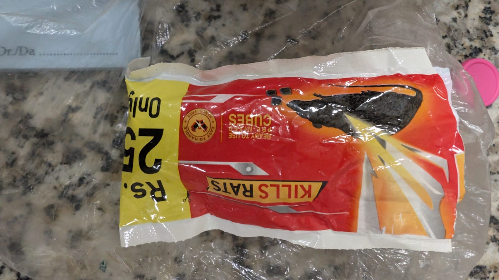
Bromadiolone poisoning is an important cause of potentially severe and prolonged bleeding because bromadiolone is a long-acting anticoagulant rodenticide (LAAR), commonly called a superwarfarin. It inhibits vitamin K recycling and consequently reduces the activity of vitamin K–dependent coagulation factors II, VII, IX and X.
Unlike ordinary warfarin exposure, bromadiolone poisoning may produce prolonged coagulopathy lasting weeks to months, and patients may require prolonged treatment with vitamin K1 (phytomenadione) and repeated INR monitoring.
Clinical pearl: A patient can appear clinically well initially and subsequently develop severe coagulopathy. A normal INR early after exposure does not necessarily exclude clinically important toxicity.
What is Bromadiolone?
Bromadiolone is a second-generation 4-hydroxycoumarin anticoagulant rodenticide. It belongs to the long-acting anticoagulant rodenticide group along with compounds such as brodifacoum and difenacoum.
It is considerably more persistent than conventional warfarin and can produce prolonged suppression of vitamin K–dependent coagulation.
Mechanism of bromadiolone poisoning
Bromadiolone inhibits vitamin K epoxide reductase, preventing regeneration of active vitamin K.
This results in impaired γ-carboxylation and activation of:
Factor II
Factor VII
Factor IX
Factor X
Protein C
Protein S
The result is progressive impairment of coagulation and an increased risk of spontaneous or trauma-related bleeding.
Clinical Features of Bromadiolone Poisoning
Patients may initially have no symptoms.
As coagulopathy develops, manifestations may include:
Epistaxis
Gingival bleeding
Easy bruising
Hematuria
Hematemesis
Melena
Hematochezia
Menorrhagia
Intramuscular bleeding
Hemarthrosis
Retroperitoneal bleeding
Intracranial hemorrhage
Hemoperitoneum
Severe anemia
Hemorrhagic shock
Intracranial hemorrhage is an uncommon but potentially life-threatening complication.
Initial Assessment
Management begins with stabilization and assessment of bleeding risk.
ABCDE assessment
A — Airway
Assess airway protection, particularly in patients with:
Altered consciousness
Intracranial hemorrhage
Massive upper gastrointestinal bleeding
B — Breathing
Assess:
Respiratory rate
Oxygen saturation
Respiratory distress
C — Circulation
Assess:
Heart rate
Blood pressure
Peripheral perfusion
Capillary refill
Evidence of active bleeding
Shock
Establish IV access and obtain blood samples early.
D — Disability
Assess:
Glasgow Coma Scale
Pupils
Focal neurological deficits
Headache
Seizures
Consider intracranial hemorrhage when neurological symptoms occur in a patient with severe coagulopathy.
E — Exposure
Look carefully for:
Bruising
Petechiae
Hematomas
Hematuria
Gastrointestinal bleeding
Injection or trauma sites
Investigations in Bromadiolone Poisoning
Important investigations include:
Essential tests
PT/INR
aPTT
CBC with platelet count
Hemoglobin/hematocrit
Blood group and crossmatch
Fibrinogen
Renal function
Liver function tests
Serum electrolytes
Additional tests when indicated
Factor II, VII, IX and X levels
Mixing study
Specific bromadiolone/superwarfarin assay
CT brain for neurological symptoms
Ultrasound/CT for suspected internal bleeding
Bromadiolone poisoning characteristically produces a vitamin K–dependent factor deficiency pattern, particularly involving factors II, VII, IX and X.
Bromadiolone Poisoning Treatment
The two major principles of treatment are:
Replace vitamin K1 to overcome the anticoagulant effect
Rapidly replace deficient coagulation factors when there is significant or life-threatening bleeding
1. Vitamin K1 — The Specific Antidote
Vitamin K1 (phytomenadione/phytonadione) is the cornerstone of treatment.
It restores the availability of reduced vitamin K and allows synthesis of functional vitamin K–dependent clotting factors.
Importantly, vitamin K does not immediately replace circulating clotting factors. Therefore, a patient with life-threatening hemorrhage may require coagulation-factor replacement in addition to vitamin K.
Route of vitamin K1
Depending on severity:
Oral vitamin K1 — useful for stable patients and prolonged treatment
IV vitamin K1 — preferred when rapid treatment is required or oral administration is not feasible
IV vitamin K should be administered cautiously because serious hypersensitivity/anaphylactoid reactions have been reported.
Vitamin K1 Dose in Bromadiolone Poisoning
There is no universally standardized dose or duration for bromadiolone poisoning.
The required dose depends on:
INR
Severity of coagulopathy
Presence or absence of bleeding
Amount/type of rodenticide exposure
Response to treatment
Recurrence of coagulopathy after dose reduction or interruption
Published cases have used a wide range of regimens, including repeated IV vitamin K followed by high-dose oral vitamin K.
For severe superwarfarin poisoning, high-dose vitamin K1 administered repeatedly throughout the day may be necessary, with dosing adjusted according to the INR and clinical response. Some toxicology literature reports doses as high as 50–100 mg/day or more in severe cases, but these doses should be individualized with toxicology/hematology guidance rather than applied as a routine dose.
Important
Do not use a single fixed vitamin K dose for every bromadiolone ingestion.
and can rapidly correct severe vitamin K antagonist–associated coagulopathy.
Fresh Frozen Plasma
FFP can also replace deficient coagulation factors.
It may be used when PCC is unavailable or according to local protocol.
Packed RBCs
Packed red blood cells should be administered when significant blood loss has caused clinically important anemia or hemorrhagic shock.
The choice and dose of blood products should be individualized according to bleeding severity, hemoglobin, hemodynamic status and local transfusion protocols.
Key principle
Major bleeding = vitamin K1 + rapid coagulation-factor replacement + definitive control of the bleeding source.
3. Control the Source of Bleeding
Correction of coagulopathy is not enough if active bleeding continues.
Depending on the site, management may include:
Endoscopic hemostasis
Surgical intervention
Interventional radiology
Neurosurgical intervention
Local pressure
Gynecological intervention
Management of gastrointestinal bleeding
A published case of severe bromadiolone-associated intracranial/other hemorrhage illustrates the need for both correction of coagulopathy and definitive treatment of the bleeding source.
4. Activated Charcoal and Gastric Decontamination
Routine gastrointestinal decontamination is not the main treatment for established bromadiolone poisoning.
Evidence summarized in toxicology reviews indicates that multiple-dose activated charcoal has not demonstrated reliable benefit for superwarfarin poisoning.
Any consideration of activated charcoal after a recent ingestion should therefore be individualized according to:
Time since ingestion
Amount ingested
Airway protection
Toxicology advice
Product formulation
Do not delay resuscitation or treatment of hemorrhage for gastrointestinal decontamination.
5. INR Monitoring
INR is the most practical laboratory marker for monitoring the anticoagulant effect.
A patient with significant exposure should have serial:
PT/INR
aPTT
CBC
Hemoglobin
The frequency depends on the severity of poisoning and treatment response.
In severe poisoning, INR may need to be checked frequently during initial stabilization, followed by less frequent monitoring once a stable vitamin K regimen has been established.
Why Long-Term Treatment May Be Necessary
This is one of the most important differences between bromadiolone and ordinary warfarin poisoning.
Bromadiolone is highly lipophilic and has prolonged persistence in the body. Human pharmacokinetic data demonstrate a prolonged terminal elimination phase; one clinical study estimated a terminal half-life of approximately 24 days.
Therefore:
Stopping vitamin K too early → recurrent INR elevation → recurrent bleeding.
Cases have documented recurrence of severe coagulopathy after vitamin K was discontinued despite an initially normal INR.
When Can Vitamin K Be Stopped?
There is no universally accepted evidence-based stopping rule for bromadiolone poisoning.
A normal INR while the patient is receiving vitamin K does not necessarily mean that bromadiolone has been eliminated.
A commonly used approach is:
Stabilize the INR with vitamin K.
Continue vitamin K while the toxic effect persists.
Gradually reduce/taper treatment when appropriate.
Stop vitamin K under specialist supervision.
Recheck PT/INR approximately 48–72 hours after stopping vitamin K.
Restart treatment if significant coagulopathy recurs.
Where available, quantitative serum bromadiolone/superwarfarin testing can provide additional information, although there is no universally validated concentration threshold that independently determines when treatment can safely stop.
Obtain PT/INR ± aPTT, CBC and other baseline investigations
↓
Is there major/life-threatening bleeding?
YES
→ Vitamin K1 → 4-factor PCC or FFP for rapid factor replacement → RBC transfusion when indicated → Definitive control of bleeding → Serial INR monitoring → Toxicology/hematology consultation
NO
→ Assess exposure and coagulation profile → Serial PT/INR monitoring → Vitamin K1 when clinically indicated → Continue monitoring because delayed/prolonged coagulopathy can occur
→ Check adherence → Reassess ongoing exposure/re-exposure → Increase/adjust vitamin K under specialist guidance → Evaluate for ongoing bleeding
↓
Considering stopping vitamin K?
→ Stop only after adequate clinical assessment → Recheck INR after approximately 48–72 hours → Restart treatment if coagulopathy recurs
Bromadiolone Poisoning in Children
Children may accidentally ingest rodenticide bait.
A child who has ingested a small amount may remain asymptomatic, but the risk assessment depends on:
Exact product
Active ingredient
Concentration
Amount ingested
Child’s weight
Time since ingestion
Repeated versus single exposure
Do not automatically assume that every rodenticide product contains bromadiolone. The product label should be checked whenever possible.
For children with suspected anticoagulant rodenticide exposure, consultation with a poison center or medical toxicologist is recommended.
Important Differential Diagnoses
A markedly prolonged PT/INR should not automatically be attributed to bromadiolone.
Consider:
Warfarin exposure
Other anticoagulant rodenticides
Vitamin K deficiency
Liver disease
Disseminated intravascular coagulation
Acquired factor inhibitors
Congenital coagulation-factor deficiencies
Malabsorption
Drug interactions
Superwarfarin poisoning should particularly be considered in a patient with unexplained prolonged PT/INR and bleeding, especially when the history is unclear.
Key Differences: Warfarin vs Bromadiolone
Feature
Warfarin
Bromadiolone
Use
Therapeutic anticoagulant
Rodenticide
Class
Vitamin K antagonist
Long-acting vitamin K antagonist
Common name
Warfarin
Superwarfarin
Duration of effect
Usually shorter
Prolonged
Coagulopathy
Usually manageable over days
May persist weeks–months
Antidote
Vitamin K1
Vitamin K1
Severe bleeding
Factor replacement may be required
Factor replacement may be required
Long-term vitamin K
Sometimes
Frequently required in severe poisoning
Monitoring
INR
Serial INR/PT
Important Clinical Pearls
1. A normal early INR does not completely exclude toxicity.
The anticoagulant effect can be delayed because existing circulating clotting factors must first decline.
2. Vitamin K1 is the specific treatment.
Bromadiolone poisoning is fundamentally a vitamin K antagonist poisoning.
3. Vitamin K does not immediately correct major hemorrhage.
In life-threatening bleeding, rapidly replace coagulation factors with 4-factor PCC or FFP, according to availability and local protocol.
4. Treatment may last for months.
Severe bromadiolone poisoning can require prolonged vitamin K1 therapy because of its long persistence.
5. Do not stop vitamin K simply because the INR becomes normal.
The INR may normalize because of administered vitamin K while the rodenticide remains in the body.
6. Monitor after stopping therapy.
A recurrence of INR elevation after vitamin K withdrawal strongly suggests persistent anticoagulant activity.
7. Always identify the active ingredient.
“Rat poison” is not a diagnosis. Different rodenticides have different toxic mechanisms and treatments.
Summary
Bromadiolone poisoning is a potentially life-threatening superwarfarin poisoning characterized by prolonged vitamin K–dependent coagulopathy.
The cornerstone of treatment is:
Vitamin K1 + INR-guided monitoring + rapid factor replacement when significant bleeding is present.
Patients with severe poisoning may require high-dose and prolonged vitamin K1 therapy for weeks or months. Treatment should not be discontinued solely because the INR has normalized while the patient is receiving vitamin K. After vitamin K is eventually stopped, repeat coagulation testing after approximately 48–72 hours can help identify recurrent anticoagulation.
Because bromadiolone poisoning can be prolonged and potentially fatal, significant exposures, abnormal coagulation studies, or any active bleeding warrant urgent medical evaluation and specialist toxicology/hematology input.
Frequently Asked Questions
What is the antidote for bromadiolone poisoning?
Vitamin K1 (phytomenadione/phytonadione) is the specific antidote for bromadiolone-induced vitamin K antagonism.
How long does bromadiolone poisoning last?
Severe poisoning can persist for weeks to months, reflecting the prolonged persistence of bromadiolone and its anticoagulant effect.
Does bromadiolone poisoning cause bleeding?
Yes. Severe poisoning can cause epistaxis, gum bleeding, hematuria, gastrointestinal bleeding, internal hemorrhage and, rarely, intracranial hemorrhage.
Can bromadiolone poisoning be treated?
Yes. Early recognition, vitamin K1 therapy, appropriate coagulation-factor replacement and close monitoring can successfully reverse the coagulopathy.
Is vitamin K enough for severe bleeding?
Not necessarily. Vitamin K restores coagulation factor production but does not provide an immediate supply of functional factors. Patients with life-threatening bleeding may require 4-factor PCC or FFP in addition to vitamin K1.
Can bromadiolone poisoning recur after treatment?
Yes. Recurrent coagulopathy can occur after vitamin K is stopped because bromadiolone may remain active in the body for a prolonged period.
The Direct Coombs Test (DCT), also known as the Direct Antiglobulin Test (DAT), is an important laboratory investigation used to detect immunoglobulins or complement components attached to the surface of red blood cells (RBCs). It is particularly useful in the evaluation of suspected immune-mediated hemolysis, autoimmune hemolytic anemia, hemolytic disease of the fetus and newborn, and hemolytic transfusion reactions.
This article explains the principle of the Direct Coombs Test, the major clinical conditions in which it is used, and how to interpret its results in pediatric and neonatal practice.
What Is the Direct Coombs Test?
The Direct Coombs Test detects IgG antibodies and/or complement components attached to the patient’s red blood cells in vivo. These substances may be responsible for immune-mediated destruction of RBCs.
In the laboratory, the patient’s RBCs are washed to remove unbound proteins. Antihuman globulin reagent is then added. If immunoglobulin or complement is attached to the RBC surface, the reagent may cause visible agglutination, resulting in a positive test.
Principle of the Direct Antiglobulin Test
Red blood cells are collected from the patient.
The RBCs are washed to remove unbound antibodies and plasma proteins.
Antihuman globulin reagent is added.
Agglutination indicates that immunoglobulin and/or complement is attached to the RBCs.
A positive DCT indicates that RBCs are coated with immunoglobulin or complement. However, it does not independently establish that clinically significant hemolysis is occurring.
Conditions in Which the Direct Coombs Test Is Used
The DCT is primarily used when immune-mediated red blood cell destruction is suspected. The following are the major clinical conditions associated with its use.
1. Autoimmune Hemolytic Anemia
Autoimmune hemolytic anemia (AIHA) occurs when the immune system produces antibodies against the patient’s own red blood cells, resulting in premature RBC destruction.
The Direct Coombs Test is an important investigation in suspected AIHA because it can demonstrate immunoglobulin or complement attached to the RBC surface.
Warm autoimmune hemolytic anemia: Usually associated with IgG coating of RBCs. The DCT is commonly positive for IgG, with or without complement.
Cold agglutinin disease: Commonly associated with complement, particularly C3, attached to RBCs. The IgG component may be negative.
Clinical findings that may support a diagnosis of AIHA include pallor, jaundice, dark urine, splenomegaly, anemia, reticulocytosis, elevated lactate dehydrogenase, and reduced haptoglobin.
2. Hemolytic Disease of the Fetus and Newborn
Hemolytic disease of the fetus and newborn (HDFN) occurs when maternal IgG antibodies cross the placenta and bind to antigens on fetal or neonatal red blood cells.
The DCT is used on the newborn’s RBCs to detect maternal antibodies attached to the cells.
It is particularly relevant in cases involving:
Rh incompatibility, including maternal anti-D antibodies.
ABO incompatibility, especially when maternal anti-A or anti-B IgG antibodies affect the newborn.
Other clinically significant maternal red-cell alloantibodies, such as anti-c or anti-K.
A newborn with jaundice and anemia, especially in the presence of maternal–fetal blood-group incompatibility, may require a DCT as part of the evaluation.
Important neonatal point: A positive DCT does not necessarily mean that the newborn has clinically significant hemolysis. The result should be interpreted together with hemoglobin, bilirubin, reticulocyte count, peripheral blood smear, and the clinical condition of the baby.
3. ABO Incompatibility in Newborns
ABO incompatibility can occur when a mother has blood group O and the newborn has blood group A or B. Maternal IgG anti-A or anti-B antibodies may cross the placenta and bind to neonatal RBCs.
The DCT may be positive in an affected newborn. However, the test can be negative in some cases of clinically significant ABO hemolysis, and a positive result does not always indicate substantial hemolysis.
Therefore, the DCT should not be used in isolation to determine the severity of neonatal jaundice or to decide on treatment.
4. Rh Isoimmunization
Rh isoimmunization occurs when a mother develops antibodies against Rh antigens present on fetal red blood cells. Maternal anti-D IgG is a classic example.
When these antibodies cross the placenta, they may cause fetal or neonatal hemolysis. The Direct Coombs Test can detect maternal antibodies attached to the newborn’s RBCs.
Depending on the severity, affected newborns may develop anemia, jaundice, or, in severe cases, hydrops fetalis.
5. Hemolytic Transfusion Reactions
The Direct Coombs Test may be useful in the investigation of suspected immune-mediated hemolytic transfusion reactions.
Acute Hemolytic Transfusion Reaction
An acute hemolytic transfusion reaction may occur when incompatible blood is transfused, leading to immune-mediated destruction of transfused RBCs. The DCT may become positive when immunoglobulin or complement is attached to RBCs.
Delayed Hemolytic Transfusion Reaction
A delayed hemolytic transfusion reaction usually occurs days to weeks after a transfusion when an alloantibody-mediated immune response leads to destruction of transfused RBCs.
The DCT may be positive during the investigation, although the result depends on the antibody, the timing of testing, and the degree of RBC coating.
Evaluation should also include transfusion history, antibody screening, antibody identification, hemoglobin, bilirubin, reticulocyte count, and other appropriate hemolysis investigations.
6. Drug-Induced Immune Hemolytic Anemia
Some medications can trigger immune-mediated hemolytic anemia. Drug-induced immune hemolytic anemia may occur through different mechanisms, including drug-dependent or drug-independent antibodies.
The DCT may be positive for IgG, complement, or both, depending on the mechanism involved.
When drug-induced hemolysis is suspected, the medication history is essential. The laboratory findings must be interpreted with clinical evidence of hemolysis and, when necessary, specialized immunohematological testing.
7. Systemic Lupus Erythematosus
Systemic lupus erythematosus (SLE) and other autoimmune disorders may be associated with autoimmune hemolytic anemia.
The DCT can be positive when RBC-bound antibodies or complement are present. However, a positive DCT in a patient with SLE does not necessarily indicate active hemolysis.
The diagnosis of autoimmune hemolytic anemia requires correlation with anemia, evidence of increased RBC destruction, and exclusion of other causes.
8. Lymphoproliferative Disorders
Lymphoproliferative disorders, including certain leukemias and lymphomas, may be associated with autoimmune hemolytic anemia.
The DCT can help identify RBC-bound immunoglobulin or complement in patients with suspected immune-mediated hemolysis. The result should be interpreted alongside the patient’s clinical presentation and hematological investigations.
Direct Coombs Test: DCT vs. Indirect Coombs Test
The Direct and Indirect Coombs Tests are both antiglobulin tests, but they detect different targets.
Feature
Direct Coombs Test (DCT)
Indirect Coombs Test (ICT)
Also called
Direct Antiglobulin Test (DAT)
Indirect Antiglobulin Test (IAT)
What it detects
IgG and/or complement attached to RBCs
Antibodies present in serum or plasma that can bind to RBC antigens
Where the antibodies are detected
On the RBC surface
In the serum or plasma
Major clinical use
Immune hemolysis and HDFN evaluation
Pretransfusion antibody screening and antenatal antibody screening
Neonatal application
Detects maternal antibodies attached to neonatal RBCs
Detects clinically significant antibodies in maternal serum
How to Interpret a Positive DCT
A positive DCT means that immunoglobulin and/or complement has been detected on the surface of the tested RBCs.
A positive result may be seen in:
Autoimmune hemolytic anemia.
Hemolytic disease of the fetus and newborn.
Immune-mediated hemolytic transfusion reactions.
Drug-induced immune hemolytic anemia.
Some autoimmune and lymphoproliferative disorders.
Some patients without clinically important hemolysis.
A positive DCT is not synonymous with hemolytic anemia. The presence of RBC-bound immunoglobulin or complement must be interpreted in the context of the patient’s clinical findings and laboratory evidence.
Investigations to Correlate With DCT Results
When immune hemolysis is suspected, relevant investigations may include:
Complete blood count and hemoglobin concentration.
Reticulocyte count.
Total and indirect bilirubin.
Lactate dehydrogenase (LDH).
Haptoglobin, when appropriate.
Peripheral blood smear.
Blood group and antibody screening.
Clinical history of transfusion, medication exposure, and maternal–fetal blood-group incompatibility.
Can the Direct Coombs Test Be Negative in Hemolysis?
Yes. A negative DCT does not completely exclude immune-mediated hemolysis.
Possible explanations include:
The quantity of RBC-bound antibody is below the detection threshold of the test.
The antibody class or subtype is not adequately detected by the reagent used.
The RBC-bound immunoglobulin has been removed or reduced before testing.
The hemolysis is nonimmune in origin.
In patients with strong clinical evidence of autoimmune hemolytic anemia but a negative routine DCT, further evaluation with an appropriate laboratory may be considered.
High-Yield Clinical Summary
DCT/DAT: Detects IgG and/or complement attached to the patient’s RBCs.
Warm AIHA: Usually associated with IgG-positive RBCs.
Cold agglutinin disease: Commonly associated with C3-positive RBCs.
HDFN: DCT may detect maternal IgG attached to neonatal RBCs.
Transfusion reactions: DCT may support the evaluation of immune-mediated hemolysis.
Positive DCT: Does not automatically prove active hemolysis.
Negative DCT: Does not completely exclude immune hemolysis.
Clinical correlation: Always interpret the result alongside evidence of hemolysis and the clinical context.
Frequently Asked Questions (FAQs)
1. What is the main purpose of the Direct Coombs Test?
The main purpose of the DCT is to detect immunoglobulin or complement attached to the surface of red blood cells. It is commonly used in the evaluation of autoimmune hemolytic anemia, hemolytic disease of the fetus and newborn, and immune-mediated transfusion reactions.
2. Is the Direct Coombs Test positive in ABO incompatibility?
It may be positive when maternal IgG anti-A or anti-B antibodies bind to neonatal RBCs. However, not every newborn with ABO incompatibility has a positive DCT, and a positive result does not necessarily indicate significant hemolysis.
3. What is the difference between DCT and ICT?
The DCT detects antibodies or complement attached to RBCs, whereas the ICT detects antibodies in serum or plasma that can bind to RBC antigens. The DCT is commonly used in evaluating immune hemolysis, while the ICT is used in antibody screening and compatibility testing.
4. Does a positive DCT confirm autoimmune hemolytic anemia?
No. A positive DCT supports the presence of RBC-bound immunoglobulin or complement, but autoimmune hemolytic anemia requires evidence of hemolysis and appropriate clinical correlation.
5. Can the DCT be negative in autoimmune hemolytic anemia?
Yes. Some patients with autoimmune hemolytic anemia may have a negative routine DCT because of low levels of RBC-bound antibodies or limitations of the testing method.
Conclusion
The Direct Coombs Test (DCT) is an essential investigation for detecting immunoglobulin or complement attached to red blood cells. Its major clinical applications include autoimmune hemolytic anemia, hemolytic disease of the fetus and newborn, immune-mediated transfusion reactions, and drug-induced immune hemolysis.
For pediatricians and neonatologists, the DCT is particularly important in evaluating newborns with suspected immune-mediated hemolysis. Nevertheless, the test result should always be interpreted together with the clinical condition, hemoglobin, bilirubin, reticulocyte count, and other relevant investigations.
Educational note: This article is intended for medical education. Diagnosis and treatment should be guided by the clinical context, institutional protocols, and appropriate specialist consultation.
Neutropenia = ANC below the age-appropriate normal lower limit.
In older children/adults, a commonly used cutoff is:
ANC <1,500 cells/µL (1.5 × 10⁹/L)
However, ANC must be interpreted according to age and population, particularly in infants and young children. The infection risk correlates much more strongly with the severity and duration of neutropenia than simply with whether ANC is below 1,500. (MSD Manuals)
For pediatrics, the key is to distinguish neutropenia (a laboratory finding) from febrile neutropenia (a clinical emergency defined by fever + sufficiently low ANC).
These are the conventional severity categories used clinically. (MSD Manuals)
Important threshold
ANC <500/µL = severe neutropenia.
At this level, organisms from the patient’s own oral/GI flora can produce serious infection. At ANC <200/µL, signs of inflammation can be particularly muted. (MSD Manuals)
The distinction matters because chemotherapy-associated febrile neutropenia is a high-risk infectious syndrome, whereas many otherwise healthy children develop transient neutropenia during viral infections.
9. Management of neutropenia
A. First question: Is the child febrile or clinically unwell?
If fever + severe neutropenia
Treat as a potentially serious infection.
Immediate:
ABC/clinical stability
Full examination
CBC + differential
Blood cultures
Urine evaluation/culture when appropriate
Evaluate for focal infection
Start appropriate empiric antibiotics promptly
For high-risk pediatric oncology patients, institutional protocols commonly use an antipseudomonal β-lactam such as cefepime, with escalation/additional coverage based on instability, focal infection, resistant organisms and local epidemiology. (IDMP)
Do not wait for the ANC to recover before treating a clinically significant suspected infection.
10. If the child is afebrile
Management depends on:
ANC
Duration of neutropenia
Age
Previous infections
Underlying disease
Chemotherapy/HSCT status
Medications
Clinical appearance
Mild ANC 1,000–1,500
Usually:
Observe + repeat CBC + investigate cause if persistent.
Moderate ANC 500–1,000
Look for cause and follow trend.
Severe ANC <500
Need much closer assessment, particularly if persistent.
Profound ANC <200
Highest concern for serious infection; clinical signs may be deceptively subtle. (MSD Manuals)
11. Treat the cause
Examples:
Drug-induced → stop suspected offending drug when appropriate.
Viral → usually supportive treatment and serial ANC.
Autoimmune neutropenia → often observation if child is clinically well; treatment depends on severity/infections.
Congenital/severe chronic neutropenia → hematology evaluation; G-CSF may be indicated.
Chemotherapy-induced → oncology-directed management ± G-CSF depending on regimen/risk.
Bone marrow failure/infiltration → urgent hematology evaluation.
G-CSF is particularly useful in selected severe/chronic neutropenia and in prevention/reduction of chemotherapy-related neutropenia. (MSD Manuals)
High-yield exam summary
Neutropenia = low ANC.
ANC:
1000–1500 → mild
500–1000 → moderate
<500 → severe
<200 → profound
Febrile neutropenia:
Fever ≥38.3°C once OR ≥38.0°C for ≥1 h + ANC <500/µL (or expected to fall <500 within 48 h).
And remember:
ANC <500 without fever = severe neutropenia, NOT febrile neutropenia.
Fever with ANC 500–1000 = febrile illness with moderate neutropenia, not the conventional febrile-neutropenia definition.
The classic “febrile neutropenia” pathway is particularly relevant to children receiving chemotherapy/HSCT; don’t automatically equate every viral-associated neutropenia in a healthy child with chemotherapy-associated FN. (IDMP)
Vitamin-responsive seizures are uncommon, but they represent some of the most important treatable causes of neonatal and early-infantile epilepsy. Recognizing them early can prevent status epilepticus, developmental injury, and potentially irreversible neurological damage.
Introduction
Neonatal seizures are usually approached as an emergency, with hypoxic-ischemic injury, hypoglycemia, hypocalcemia, intracranial hemorrhage, stroke, infection, and structural brain abnormalities among the major considerations.
However, a small but particularly important group of seizures results from inborn errors of metabolism involving vitamins or vitamin-dependent enzymes. These seizures may be remarkably resistant to conventional antiseizure medications but respond dramatically to administration of the appropriate vitamin or cofactor.
The major clinically relevant disorders include:
Pyridoxine-dependent epilepsy (PDE)
Pyridoxal-5′-phosphate (PLP)-dependent epilepsy
Folinic acid-responsive seizures
Biotinidase deficiency
Biotin-thiamine-responsive basal ganglia disease
Other metabolic disorders in which vitamin/cofactor therapy can be seizure-modifying
Among these, vitamin B6-dependent epilepsies are particularly important in the neonate.
1. When should you suspect a vitamin-responsive seizure disorder?
The diagnosis should enter the differential particularly when a neonate or infant has:
Red flags
Seizures beginning in the neonatal period without an obvious cause
Frequent or recurrent seizures despite appropriate antiseizure medication
Status epilepticus
Abnormal EEG with burst suppression or marked discontinuity
Seizures accompanied by unexplained encephalopathy
Irritability, abnormal crying, vomiting, or feeding difficulty
Unexplained metabolic acidosis or elevated lactate
Seizures recurring after apparently successful treatment
A previous sibling who died from unexplained neonatal seizures
Consanguinity or a family history suggestive of an autosomal-recessive disorder
Seizures with unusual movements, spasms, myoclonus, or autonomic features
Seizures that respond dramatically to pyridoxine or PLP
The ILAE specifically recommends considering a trial of pyridoxine in neonates with clinical features suggestive of vitamin B6-dependent epilepsy and in neonates whose seizures remain unexplained and refractory to second-line antiseizure medication.
2. Pyridoxine-dependent epilepsy
The classic vitamin-responsive epilepsy
Pyridoxine-dependent epilepsy is one of the most important treatable causes of neonatal-onset epilepsy.
The classical form is caused by pathogenic variants in ALDH7A1, which encodes antiquitin, an enzyme involved in lysine degradation. Deficiency results in accumulation of metabolites including α-aminoadipic semialdehyde (α-AASA)/P6C. P6C can inactivate pyridoxal-5′-phosphate (PLP), ultimately impairing neurotransmitter synthesis and producing seizures.
Clinical presentation
The classical presentation is:
A neonate with otherwise unexplained, frequent, treatment-resistant seizures.
Seizures may be:
Focal
Multifocal
Generalized
Clonic
Tonic
Myoclonic
Epileptic spasms
Some infants may initially appear encephalopathic rather than having obvious convulsive seizures.
The ILAE recognizes frequent drug-resistant seizures, often progressing to status epilepticus, with marked reduction or cessation following pyridoxine supplementation as characteristic of vitamin B6-dependent epilepsy.
An important clue
Some infants may have:
Irritability
Restlessness
Abnormal crying
Vomiting
Feeding difficulties
before or around the time of seizures.
3. Why does pyridoxine stop the seizure?
Vitamin B6 exists in several biologically active forms. Pyridoxal-5′-phosphate (PLP) is the major active cofactor involved in numerous enzymatic reactions.
One of its critical neurological roles is participation in neurotransmitter synthesis.
In pyridoxine-dependent epilepsy, abnormal lysine metabolism leads to accumulation of metabolites that interfere with PLP availability.
Therefore:
ALDH7A1 defect
↓
Abnormal lysine degradation
↓
↑ α-AASA/P6C
↓
PLP inactivation
↓
Impaired neurotransmitter synthesis
↓
Neuronal hyperexcitability
↓
Seizures
Administration of pharmacological doses of pyridoxine restores adequate PLP availability and suppresses seizures.
4. Pyridoxine trial
This is one of the most important practical points in neonatal neurology.
A pyridoxine trial should be performed under continuous cardiorespiratory and preferably EEG monitoring, because intravenous pyridoxine can cause significant apnea, respiratory depression, and prolonged somnolence.
For prolonged/refractory clinical seizures, a commonly used approach is:
Pyridoxine 100 mg IV, with EEG and cardiorespiratory monitoring.
If there is no response, further doses may be administered according to specialist/neonatal protocol, with some references allowing repeated dosing up to a cumulative 500 mg.
The ILAE notes that responses can occasionally be delayed; therefore, lack of an immediate response does not automatically exclude vitamin B6-dependent epilepsy. A trial may need to be continued for several days in selected patients.
Important safety point
Do not give a high-dose IV pyridoxine trial to an unsupervised neonate.
Apnea and cardiorespiratory depression can occur, so airway and ventilatory support must be immediately available.
5. Pyridoxine maintenance therapy
Once pyridoxine-dependent epilepsy is established, treatment is generally lifelong.
Current International PDE Consortium recommendations summarized by GeneReviews include:
Age
Pyridoxine dose
Newborn
100 mg/day
Infant
30 mg/kg/day, maximum 300 mg/day
Child/adolescent/adult
30 mg/kg/day, maximum 500 mg/day
Doses should be individualized by a metabolic/pediatric neurology team.
Long-term excessive pyridoxine can produce sensory neuropathy, so high-dose therapy requires monitoring.
6. Pyridoxal-5′-phosphate-dependent epilepsy
Not every vitamin B6-responsive seizure disorder responds to pyridoxine itself.
This distinction is crucial.
Pyridoxamine-5′-phosphate oxidase (PNPO) deficiency results from pathogenic variants in PNPO.
The enzyme is required for conversion of pyridoxine/pyridoxamine derivatives to the active cofactor PLP.
Therefore:
Pyridoxine → requires metabolic activation → PLP
If this pathway is defective, administering pyridoxine may not adequately correct the biochemical defect.
These patients may instead respond to pyridoxal-5′-phosphate (PLP).
Practical distinction
Disorder
Main defect
Effective vitamin form
Pyridoxine-dependent epilepsy
ALDH7A1/antiquitin pathway
Pyridoxine (B6)
PNPO deficiency
Conversion to active PLP impaired
Pyridoxal-5′-phosphate
PLPBP deficiency
PLP binding/transport-related dysfunction
Often B6/PLP-dependent
The ILAE recognizes ALDH7A1, PNPO and PLPBP-related disorders among the genetic causes of vitamin B6-dependent epilepsy.
Deficiency can produce neonatal or infantile epilepsy with variable response to pyridoxine or PLP.
This disorder highlights an important clinical principle:
A negative response to one form of vitamin B6 does not necessarily exclude a vitamin B6-dependent epilepsy.
Patients may require a carefully monitored trial of the appropriate B6 formulation under metabolic/neurology supervision.
8. Folinic acid-responsive seizures
Another important treatable cause is folinic acid-responsive epilepsy.
These infants can present with:
Neonatal seizures
Drug-resistant epilepsy
Encephalopathy
Abnormal EEG
Sometimes a transient or incomplete response to pyridoxine
An important clinical clue is:
Seizures initially improve with pyridoxine but subsequently recur.
Folinic acid may then produce seizure control.
Interestingly, subsequent biochemical and genetic work demonstrated substantial overlap between classical folinic acid-responsive seizures and ALDH7A1-related pyridoxine-dependent epilepsy.
Thus, the old concept of completely separate “pyridoxine-responsive” and “folinic-acid-responsive” disorders has become more nuanced.
9. When should folinic acid be considered?
Folinic acid should be considered particularly when:
Neonatal seizures are unexplained
Pyridoxine produces only a partial/transient response
Seizures recur despite pyridoxine
There is biochemical/genetic evidence suggesting a folate-related disorder
Some literature describes folinic acid doses in the range of approximately 3–5 mg/kg/day, but dosing should be directed by a pediatric neurologist/metabolic specialist because the optimal regimen depends on the suspected disorder.
10. Biotin-responsive seizures
Biotinidase deficiency is another important treatable metabolic disorder.
Biotinidase is responsible for recycling biotin, an essential cofactor for several carboxylases.
Untreated profound biotinidase deficiency can produce:
Seizures
Hypotonia
Developmental delay
Ataxia
Alopecia
Eczematous rash
Conjunctivitis
Hearing loss
Optic atrophy
Respiratory abnormalities
Seizures may be focal, generalized, or myoclonic, and infantile spasms have also been reported.
The key point
This is a highly treatable disorder.
Early treatment can prevent neurological injury, and seizures can resolve rapidly after biotin supplementation.
11. Biotinidase deficiency: treatment
Current GeneReviews recommendations are:
Profound deficiency: oral biotin 5–10 mg/day
Partial deficiency: approximately 2.5–10 mg/day
Treatment is generally lifelong.
An important practical issue is that biotin can interfere with certain laboratory immunoassays, potentially producing misleading results. Clinicians should inform the laboratory when a patient is receiving pharmacological doses of biotin.
This is a particularly fascinating vitamin-responsive neurological disorder.
It results from pathogenic variants in SLC19A3, which encodes thiamine transporter 2.
It can present in three broad patterns:
Early-infantile disease
Usually before 3 months of age:
Vomiting
Feeding difficulty
Encephalopathy
Hypotonia
Seizures/infantile spasms
Respiratory failure
Severe lactic acidosis
Childhood disease
Usually presents with recurrent episodes of:
Encephalopathy
Seizures
Ataxia
Dystonia
Ophthalmoplegia
Dysphagia
Episodes may be precipitated by febrile illness or metabolic stress.
MRI clue
Symmetrical abnormalities involving the:
Caudate
Putamen
Medial thalami
with possible extension to the brainstem and other structures should raise suspicion.
13. Treatment of biotin-thiamine-responsive basal ganglia disease
Treatment should be started promptly when the disorder is suspected.
GeneReviews recommends:
Biotin: 5–10 mg/kg/day
plus
Thiamine: up to 40 mg/kg/day, maximum 1,500 mg/day
with lifelong therapy. During acute decompensation, thiamine may be increased and administered intravenously.
Early treatment is critical because neurological damage may become irreversible.
14. Other metabolic disorders where vitamins matter
Vitamin-responsive seizures should not be interpreted as only a “B6 problem.”
Other treatable metabolic epilepsies include disorders involving:
Folate metabolism
Thiamine metabolism
Biotin metabolism
Riboflavin-related pathways
Vitamin B12/cobalamin metabolism
Serine synthesis
Creatine metabolism
Glucose transport
Mitochondrial metabolism
Some are better classified as cofactor-responsive metabolic epilepsies rather than classical vitamin-dependent epilepsy.
A broad metabolic approach is therefore essential in unexplained refractory neonatal epilepsy. Reviews of inborn errors of metabolism in pediatric epilepsy emphasize pyridoxine/PLP, folinic acid and biotin among the particularly important treatable pathways.
15. Practical approach to a neonate with refractory seizures
A useful bedside approach is:
Step 1 — Stabilize
ABC + glucose
Check:
Blood glucose
Calcium
Magnesium
Sodium
Potassium
Blood gas
Lactate
Treat immediately reversible abnormalities.
Step 2 — Search for common causes
Consider:
HIE
Intracranial hemorrhage
Stroke
CNS infection
Hypoglycemia
Hypocalcemia
Electrolyte abnormalities
Structural brain disease
Step 3 — EEG
Use continuous EEG where available.
Vitamin-dependent epilepsies can have distinctive EEG abnormalities, including burst suppression or marked discontinuity, although these findings are not diagnostic by themselves.
Step 4 — If seizures remain unexplained/refractory
Consider folinic acid and other metabolic/cofactor-responsive disorders
↓
Confirm with biochemical/genetic testing
This is particularly important because the treatment is relatively simple compared with the potential consequences of missing the diagnosis. The ILAE specifically recommends a pyridoxine trial in selected neonates with unexplained refractory seizures.
18. Take-home points
Vitamin-responsive epilepsies are rare but potentially dramatically treatable causes of neonatal and infantile seizures.
Pyridoxine-dependent epilepsy is the classic disorder to remember.
ALDH7A1/antiquitin deficiency is the major cause of classical pyridoxine-dependent epilepsy.
PNPO deficiency may respond to PLP rather than pyridoxine.
A transient or incomplete pyridoxine response should not end the investigation.
Folinic acid-responsive seizures should be considered in selected refractory cases.
Biotinidase deficiency can cause seizures, hypotonia, rash, alopecia, and developmental problems—and is highly treatable.
SLC19A3-related biotin-thiamine-responsive basal ganglia disease is another important treatable disorder, particularly when encephalopathy, lactic acidosis and basal ganglia MRI abnormalities coexist.
High-dose IV pyridoxine can cause apnea and cardiorespiratory depression; administration requires appropriate monitoring and resuscitation capability.
Do not wait for genetic confirmation before treating a strongly suspected vitamin-dependent epilepsy.
References
Major pediatric/neonatal textbooks
Kliegman RM, St Geme JW, Blum NJ, Shah SS, Tasker RC, Wilson KM, eds.Nelson Textbook of Pediatrics. 22nd ed. Elsevier; 2023. Sections on neonatal seizures, epilepsy, and inborn errors of metabolism.
Eichenwald EC, Hansen AR, Martin CR, Stark AR, eds.Cloherty and Stark’s Manual of Neonatal Care. 9th ed. Wolters Kluwer; 2022. Sections on neonatal seizures and metabolic disorders.
Gleason CA, Juul SE, eds.Avery’s Diseases of the Newborn. 11th ed. Elsevier; 2024. Sections on neonatal neurologic disorders, seizures, and inborn errors of metabolism.
Volpe JJ.Volpe’s Neurology of the Newborn. 7th ed. Elsevier; 2018. Chapters on neonatal seizures and metabolic/epileptic disorders.
Guidelines and primary/review literature
Pressler RM, Cilio MR, Mizrahi EM, et al. Treatment of seizures in the neonate: Guidelines and consensus-based recommendations—Special report from the ILAE Task Force on Neonatal Seizures. Epilepsia. 2023.
Stockler S, Plecko B, Gospe SM Jr, et al. Neonatal vitamin-responsive epileptic encephalopathies. Neuropediatrics. 2011.
Coughlin CR, Tseng LA, et al. International consensus recommendations for the diagnosis and management of pyridoxine-dependent epilepsy. 2021.
GeneReviews. Pyridoxine-Dependent Epilepsy – ALDH7A1. University of Washington, Seattle. Updated 2026.
GeneReviews. Biotinidase Deficiency. University of Washington, Seattle. Updated 2026.
GeneReviews. Biotin-Thiamine-Responsive Basal Ganglia Disease. University of Washington, Seattle. Updated 2025.
Gallagher RC, Van Hove JLK, Scharer G, et al. Folinic acid-responsive seizures are identical to pyridoxine-dependent epilepsy. Ann Neurol. 2009;65(5):550-556.
Inborn Errors of Metabolism in Pediatric Epilepsy. Pediatr Neurol. Review of vitamin/cofactor-responsive epilepsies.
ILAE Task Force. Classification and diagnostic criteria for early-onset vitamin-dependent developmental and epileptic encephalopathies.
Editorial note
For a medical blog, I would not present the dosing section as a general prescription protocol. High-dose pyridoxine and PLP trials—particularly IV administration in neonates—should be performed in a monitored setting with respiratory support available. The exact treatment protocol should follow the neonatal unit’s guideline and pediatric neurology/metabolic specialist recommendations. The ILAE evidence base also acknowledges that much of the evidence for neonatal pyridoxine/PLP therapy is retrospective rather than from randomized trials.
The people who still navigate by feel and memory aren’t being stubborn. They’re preserving a relationship with the physical world that the rest of us quietly outsourced — and only now are we beginning to understand the cost.
Table of Contents
Introduction
You probably know someone like this. Ask them for directions, and they don’t reach for their phone. Instead they look briefly into the middle distance and begin: turn left out of the car park, follow the road until you pass the old brewery, keep the river on your right… They describe the world in landmarks, relationships, textures. The route lives inside them. To anyone born after 1990, watching this performance feels vaguely archaeological — like watching someone whittle a tool from flint.
The temptation is to read it as stubbornness. These are the holdouts, the refuseniks, the people who also probably still check the paper for TV listings. But psychology and neuroscience increasingly suggest that this framing is exactly backwards. The adults who still navigate by memory are not the ones who failed to adapt. They are the ones who, without necessarily meaning to, kept something the rest of us gave away — and the giving away, it turns out, cost more than we understood at the time.
What the brain was doing before we had a blue dot
When you navigate without assistance, your brain is not simply “remembering a route.” It is constructing and continuously updating what neuroscientists call a cognitive map — a relational model of your environment, built in the hippocampus, that allows you to know where you are relative to everywhere else you’ve been. It’s the mental architecture that lets you take a shortcut you’ve never consciously walked, or recover from a missed turn without dissolving into panic. Research published in Scientific Reports has confirmed that this strategy depends critically on the hippocampus, the same region deeply involved in episodic memory and our sense of personal history.
The map is not given to you. It is earned, slowly, through use. Every walk through a new neighbourhood without looking at your phone is a small act of hippocampal construction. Over years, the result is an intimate, structural knowledge of a place — the kind that older Londoners, New Yorkers, or Mumbaikars describe when they say they “know” their city. Not a list of addresses. A felt sense of how everything relates.
Navigating without assistance is not a minor quirk of habit. It is an act of cognitive maintenance — one that keeps an entire architectural system of the brain actively in use.
The trade we made, and what it did to the brain
Here is the uncomfortable part. The brain does not only have one navigation system. Alongside the hippocampal cognitive-map strategy, there is a second system, centred on the caudate nucleus, that operates through sequential motor instructions. Turn left here. In 200 metres, turn right. This system does not build a map. It does not need to know where it is. It only needs to know what to do next.
GPS, structurally, is a perfect match for the caudate system. And the brain, being ruthlessly efficient, will defer to the simpler system whenever it is available. The cumulative effect of years of GPS use, researchers found, is a measurable shift away from hippocampal navigation and toward this more passive, reactive mode. Worse, the shift is not neutral: people who used GPS more heavily over time showed steeper declines in hippocampal-dependent spatial memory. Not because they were worse at navigation to begin with, but because they had stopped asking their hippocampus to do the work.
A longitudinal study published in Scientific Reports found that greater GPS reliance over time was specifically associated with decline in hippocampal spatial memory — not merely correlated with pre-existing poor navigation ability. The decline followed the behaviour, not the other way around.
A longitudinal study published in Scientific Reports
This is the quiet exchange at the heart of the GPS era. We did not simply gain a more reliable way to find parking. We also, gradually and without realising it, stopped exercising a cognitive system that was doing considerably more than helping us get around. The hippocampus’s work is not limited to navigation — it underpins episodic memory, contextual learning, and a broader sense of spatial orientation in the world. When we stopped asking it to build maps, we stopped sharpening something that matters in ways navigation alone does not capture.
What the holdouts actually have
Return, then, to the person describing the route by the river and the brewery. They are not simply demonstrating an old skill. They are demonstrating an active cognitive infrastructure — one that is, in most of us, now fallow.
They know the spatial relationship between places they have never directly walked between. They carry the city’s geometry in their nervous system. When a road is closed, they reroute mentally, without recalculating. They can locate themselves after emerging from an underground station because they have, in their head, a running model of where they are in relation to everything else. These are not party tricks. They are the outputs of a cognitive map that has been continuously used and maintained.
The broader consequence of that maintenance is harder to name but easy to recognise. These people tend to feel located in a way that the rest of us, moving through cities with blue dots on glass, often do not. There is a groundedness that comes from genuinely knowing where you are — not being told, not following, but knowing. The loss of that is diffuse and private. Most people who have lost it do not know it is gone, because it was never the kind of thing the culture was tracking.
Most people who have lost their cognitive map do not know it is gone — because it was never the kind of thing the culture was tracking in the first place.
Can it be recovered?
The honest answer is: probably, with effort, in part. The hippocampus retains the capacity for spatial learning throughout adult life. The cognitive map is not permanently erased by years of GPS use — it is simply unexercised. The exercising can, in principle, resume. Navigating without assistance in familiar environments, deliberately and repeatedly, begins to rebuild it.
The difficulty is entirely practical. The phone is already in hand. The GPS is one tap away. The friction of choosing not to use it is exactly the kind of small daily friction that modern life has been redesigned, at every level, to eliminate. The GPS exists precisely because getting lost is unpleasant, expensive, and increasingly unacceptable in a world where arrival times are tracked and lateness is noticed. To choose, in that world, to navigate by feel is not simply a minor lifestyle preference. It is a decision to add deliberate friction to a frictionless system — and that is genuinely hard to sustain.
A more realistic approach for most people may be selective disengagement: putting the phone away on familiar routes, walking new neighbourhoods without GPS, choosing occasionally to get slightly lost as a form of maintenance rather than failure. Not an ideology. A workout.
The real cost of the deal we made
The GPS is not going away, and the case here is not that it should. The navigational gains are real. Billions of hours of collective lostness have been recovered. The technology works, and the technology is useful, and none of that is in question.
What is in question is whether the rest of the deal was understood when it was made. The cognitive map that the hippocampus builds through unassisted navigation was not only a navigation tool. It was a way of being in relationship with the physical world — of knowing, rather than following; of orienting, rather than obeying. The people who still navigate by memory kept that relationship without, in most cases, deciding to. They simply never outsourced it.
They are not stubborn. They are not behind. They are, in a quiet and structural way, still in possession of something that the rest of us traded away for a blue dot — and they are probably, as they feel the river to their right and the cathedral somewhere to the south, more located in the world than we are.
Whether the rest of us can get back to something like that is an open question. But acknowledging that the trade was made, and that it cost something real, seems like a reasonable place to start.
Based on research reported by Space Daily, May 2026 · Written with reference to Scientific Reports longitudinal spatial memory studies