The people who still navigate by feel and memory aren’t being stubborn. They’re preserving a relationship with the physical world that the rest of us quietly outsourced — and only now are we beginning to understand the cost.
Table of Contents
Introduction
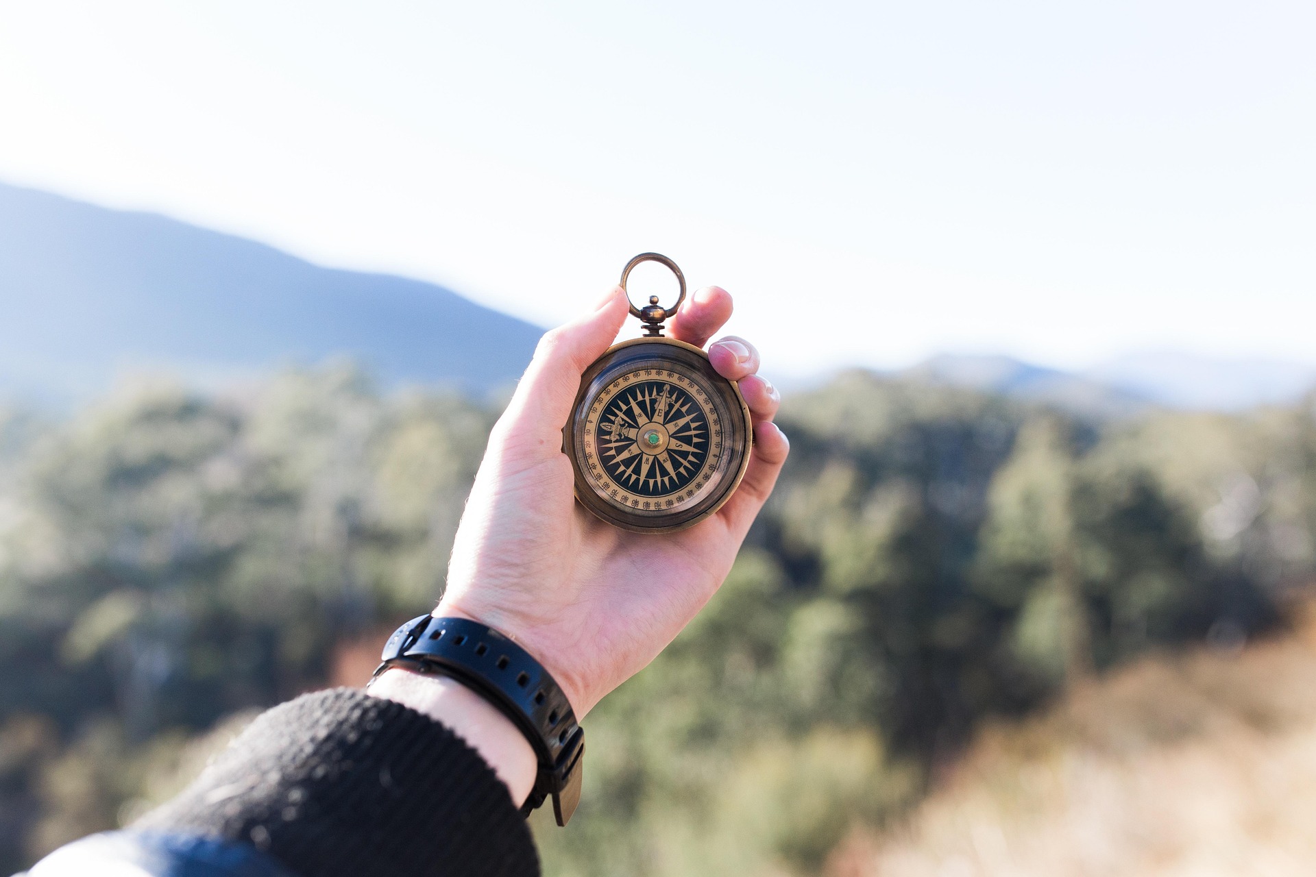
You probably know someone like this. Ask them for directions, and they don’t reach for their phone. Instead they look briefly into the middle distance and begin: turn left out of the car park, follow the road until you pass the old brewery, keep the river on your right… They describe the world in landmarks, relationships, textures. The route lives inside them. To anyone born after 1990, watching this performance feels vaguely archaeological — like watching someone whittle a tool from flint.


The temptation is to read it as stubbornness. These are the holdouts, the refuseniks, the people who also probably still check the paper for TV listings. But psychology and neuroscience increasingly suggest that this framing is exactly backwards. The adults who still navigate by memory are not the ones who failed to adapt. They are the ones who, without necessarily meaning to, kept something the rest of us gave away — and the giving away, it turns out, cost more than we understood at the time.
What the brain was doing before we had a blue dot
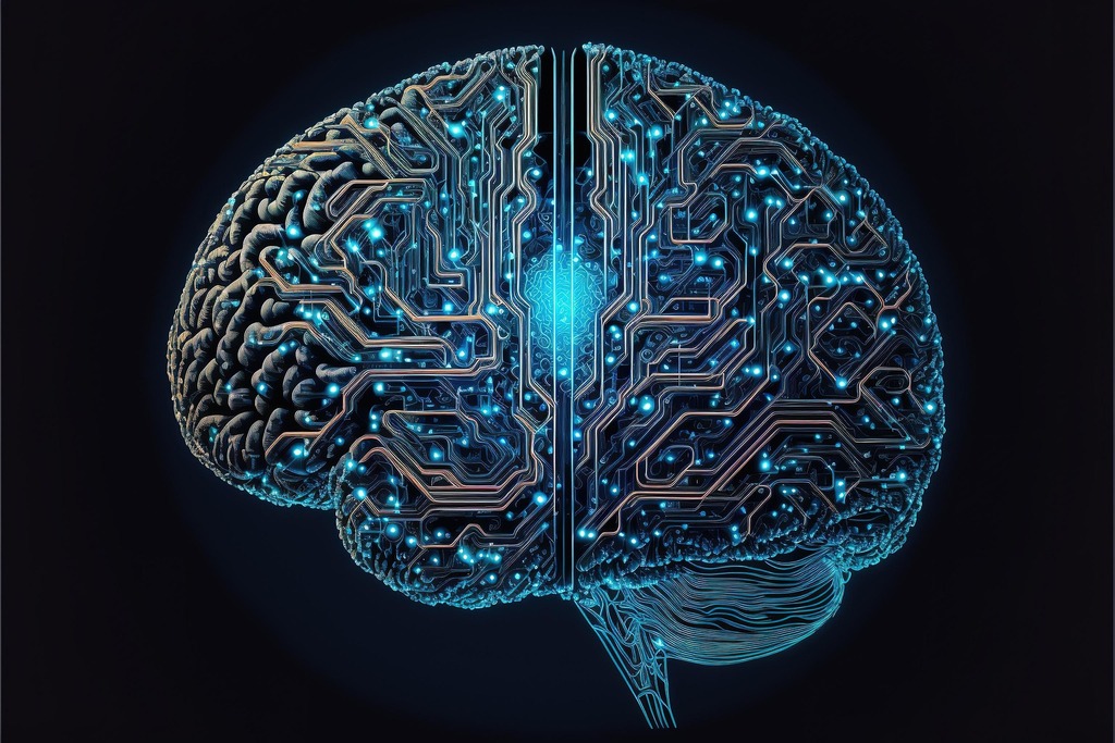
When you navigate without assistance, your brain is not simply “remembering a route.” It is constructing and continuously updating what neuroscientists call a cognitive map — a relational model of your environment, built in the hippocampus, that allows you to know where you are relative to everywhere else you’ve been. It’s the mental architecture that lets you take a shortcut you’ve never consciously walked, or recover from a missed turn without dissolving into panic. Research published in Scientific Reports has confirmed that this strategy depends critically on the hippocampus, the same region deeply involved in episodic memory and our sense of personal history.

The map is not given to you. It is earned, slowly, through use. Every walk through a new neighbourhood without looking at your phone is a small act of hippocampal construction. Over years, the result is an intimate, structural knowledge of a place — the kind that older Londoners, New Yorkers, or Mumbaikars describe when they say they “know” their city. Not a list of addresses. A felt sense of how everything relates.
Navigating without assistance is not a minor quirk of habit. It is an act of cognitive maintenance — one that keeps an entire architectural system of the brain actively in use.
The trade we made, and what it did to the brain
Here is the uncomfortable part. The brain does not only have one navigation system. Alongside the hippocampal cognitive-map strategy, there is a second system, centred on the caudate nucleus, that operates through sequential motor instructions. Turn left here. In 200 metres, turn right. This system does not build a map. It does not need to know where it is. It only needs to know what to do next.
GPS, structurally, is a perfect match for the caudate system. And the brain, being ruthlessly efficient, will defer to the simpler system whenever it is available. The cumulative effect of years of GPS use, researchers found, is a measurable shift away from hippocampal navigation and toward this more passive, reactive mode. Worse, the shift is not neutral: people who used GPS more heavily over time showed steeper declines in hippocampal-dependent spatial memory. Not because they were worse at navigation to begin with, but because they had stopped asking their hippocampus to do the work.
A longitudinal study published in Scientific Reports found that greater GPS reliance over time was specifically associated with decline in hippocampal spatial memory — not merely correlated with pre-existing poor navigation ability. The decline followed the behaviour, not the other way around.
A longitudinal study published in Scientific Reports
This is the quiet exchange at the heart of the GPS era. We did not simply gain a more reliable way to find parking. We also, gradually and without realising it, stopped exercising a cognitive system that was doing considerably more than helping us get around. The hippocampus’s work is not limited to navigation — it underpins episodic memory, contextual learning, and a broader sense of spatial orientation in the world. When we stopped asking it to build maps, we stopped sharpening something that matters in ways navigation alone does not capture.
What the holdouts actually have
Return, then, to the person describing the route by the river and the brewery. They are not simply demonstrating an old skill. They are demonstrating an active cognitive infrastructure — one that is, in most of us, now fallow.
They know the spatial relationship between places they have never directly walked between. They carry the city’s geometry in their nervous system. When a road is closed, they reroute mentally, without recalculating. They can locate themselves after emerging from an underground station because they have, in their head, a running model of where they are in relation to everything else. These are not party tricks. They are the outputs of a cognitive map that has been continuously used and maintained.
The broader consequence of that maintenance is harder to name but easy to recognise. These people tend to feel located in a way that the rest of us, moving through cities with blue dots on glass, often do not. There is a groundedness that comes from genuinely knowing where you are — not being told, not following, but knowing. The loss of that is diffuse and private. Most people who have lost it do not know it is gone, because it was never the kind of thing the culture was tracking.
Most people who have lost their cognitive map do not know it is gone — because it was never the kind of thing the culture was tracking in the first place.
Can it be recovered?
The honest answer is: probably, with effort, in part. The hippocampus retains the capacity for spatial learning throughout adult life. The cognitive map is not permanently erased by years of GPS use — it is simply unexercised. The exercising can, in principle, resume. Navigating without assistance in familiar environments, deliberately and repeatedly, begins to rebuild it.
The difficulty is entirely practical. The phone is already in hand. The GPS is one tap away. The friction of choosing not to use it is exactly the kind of small daily friction that modern life has been redesigned, at every level, to eliminate. The GPS exists precisely because getting lost is unpleasant, expensive, and increasingly unacceptable in a world where arrival times are tracked and lateness is noticed. To choose, in that world, to navigate by feel is not simply a minor lifestyle preference. It is a decision to add deliberate friction to a frictionless system — and that is genuinely hard to sustain.
A more realistic approach for most people may be selective disengagement: putting the phone away on familiar routes, walking new neighbourhoods without GPS, choosing occasionally to get slightly lost as a form of maintenance rather than failure. Not an ideology. A workout.
The real cost of the deal we made
The GPS is not going away, and the case here is not that it should. The navigational gains are real. Billions of hours of collective lostness have been recovered. The technology works, and the technology is useful, and none of that is in question.
What is in question is whether the rest of the deal was understood when it was made. The cognitive map that the hippocampus builds through unassisted navigation was not only a navigation tool. It was a way of being in relationship with the physical world — of knowing, rather than following; of orienting, rather than obeying. The people who still navigate by memory kept that relationship without, in most cases, deciding to. They simply never outsourced it.
They are not stubborn. They are not behind. They are, in a quiet and structural way, still in possession of something that the rest of us traded away for a blue dot — and they are probably, as they feel the river to their right and the cathedral somewhere to the south, more located in the world than we are.
Whether the rest of us can get back to something like that is an open question. But acknowledging that the trade was made, and that it cost something real, seems like a reasonable place to start.
Based on research reported by Space Daily, May 2026 · Written with reference to Scientific Reports longitudinal spatial memory studies