Monthly Archives: May 2026

NMCLE past questions

🩺 NMCLE Top 10 Important Past-Style Questions (with Explanations)


1. 🫀 Acute Myocardial Infarction (MI)

Q: A 55-year-old man presents with chest pain radiating to the left arm. ECG shows ST elevation in leads II, III, aVF. Which artery is most likely blocked?

Answer: Right Coronary Artery (RCA)

✔ Explanation:

  • Inferior wall MI = II, III, aVF
  • Usually due to RCA occlusion

🔥 High-yield points:

  • Anterior MI → LAD (V1–V4)
  • Lateral MI → Circumflex (I, aVL, V5–V6)
  • Inferior MI → RCA

2. 🧫 Tuberculosis Diagnosis

Q: What is the most specific test for pulmonary tuberculosis?

Answer: GeneXpert MTB/RIF

✔ Explanation:

  • Detects TB DNA + rifampicin resistance in 2 hours

🔥 High-yield:

  • Ziehl-Neelsen stain → quick but less sensitive
  • Culture (Lowenstein-Jensen) → gold standard but slow
  • GeneXpert = best for rapid diagnosis

3. 💊 Drug of choice in Anaphylaxis

Q: First-line drug in anaphylactic shock?

Answer: IM Adrenaline (Epinephrine)

✔ Explanation:

  • Acts on α1 (vasoconstriction), β1, β2 (bronchodilation)

🔥 High-yield:

  • Give in mid-thigh IM
  • Repeat every 5–15 min if needed
  • Antihistamines are NOT first-line

4. 🧠 Stroke Localization

Q: Right-sided hemiplegia with aphasia indicates lesion in?

Answer: Left middle cerebral artery (MCA)

✔ Explanation:

  • Left hemisphere = language center (Broca/Wernicke)

🔥 High-yield:

  • MCA → face & arm > leg weakness + aphasia
  • ACA → leg > arm weakness
  • PCA → visual disturbances

5. 🤰 Eclampsia Management

Q: Best drug to prevent seizures in eclampsia?

Answer: Magnesium sulfate

✔ Explanation:

  • CNS depressant stabilizes neurons

🔥 High-yield (SIPS):

  • Loading: 4g IV + 10g IM
  • Maintenance: 5g IM 4-hourly
  • Antidote: Calcium gluconate

6. 🧬 Iron Deficiency Anemia

Q: Most common cause of microcytic hypochromic anemia worldwide?

Answer: Iron deficiency anemia

✔ Explanation:

  • Due to poor intake, blood loss, malabsorption

🔥 High-yield:

  • Low ferritin = earliest marker
  • High TIBC
  • Treatment: oral ferrous sulfate

7. 🦠 HIV Diagnosis

Q: Screening test for HIV?

Answer: ELISA (or rapid antibody test)

✔ Explanation:

  • Detects antibodies to HIV

🔥 High-yield:

  • ELISA → screening
  • Western blot → confirmation (less used now)
  • PCR → early infant diagnosis

8. 👶 Neonatal Jaundice

Q: Physiological jaundice appears after how many hours?

Answer: After 24 hours of birth

✔ Explanation:

  • Due to immature liver enzymes (UGT deficiency)

🔥 High-yield:

  • Physiological:
    • Appears after 24h
    • Peaks day 3–5
  • Pathological: within 24h (always abnormal)

9. 🧪 Diabetes Mellitus Diagnosis

Q: Diagnostic fasting blood glucose for diabetes?

Answer: ≥126 mg/dL (7.0 mmol/L)

✔ Explanation:

  • Confirms impaired glucose metabolism

🔥 High-yield:

  • FBS ≥126 mg/dL
  • RBS ≥200 mg/dL + symptoms
  • HbA1c ≥6.5%

10. ⚕️ Most common site of peptic ulcer

Q: Most common site of duodenal ulcer?

Answer: First part of duodenum (bulb)

✔ Explanation:

  • Acid exposure highest here

🔥 High-yield:

  • Duodenal ulcer → pain relieved by food
  • Gastric ulcer → pain worsens with food

📌 Ultra High-Yield Revision Summary (Exam Favorites)

🚨 Must-remember lists:

  • MI leads & arteries
  • TB diagnosis hierarchy (GeneXpert > culture > smear)
  • Anaphylaxis = adrenaline
  • Eclampsia = magnesium sulfate
  • HIV = ELISA screening
  • Diabetes cutoff values
  • Neonatal jaundice timing rules

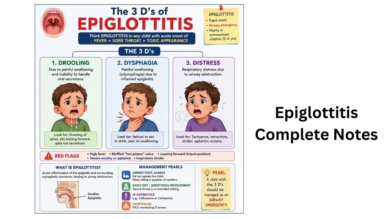
Epiglottitis – 5 clinical features, Short Notes

Definition

Epiglottitis is an acute inflammation and swelling of the epiglottis (a flap of cartilage at the base of the tongue that prevents food from entering the airway).
It is a medical emergency because swelling can rapidly block the airway. (Mayo Clinic)


Anatomy and Function of Epiglottis

  • Located above the larynx.
  • Acts like a “lid” over the trachea during swallowing.
  • Prevents aspiration of food and liquids into the lungs.

Causes

Infectious Causes

  • Haemophilus influenzae type b (Hib) – classic cause in children
  • Streptococcus pneumoniae
  • Streptococcal species
  • Staphylococcus aureus
  • Viral or fungal infections (less common)

Non-infectious Causes

  • Hot liquid burns
  • Trauma to throat
  • Chemical injury
  • Smoking/vaping or inhaling drugs (Mayo Clinic)

Risk Factors

  • Lack of Hib vaccination
  • Weak immune system
  • Diabetes
  • Smoking
  • Young children (historically), though now more common in adults

Clinical Features

Symptoms

  • Severe sore throat
  • Fever
  • Difficulty swallowing (dysphagia)
  • Painful swallowing (odynophagia)
  • Drooling
  • Muffled or “hot potato” voice
  • Difficulty breathing
  • Stridor (high-pitched breathing sound)

Signs

  • Patient sits leaning forward (tripod position)
  • Anxiety/restlessness
  • Cyanosis in severe cases

Classic Presentation

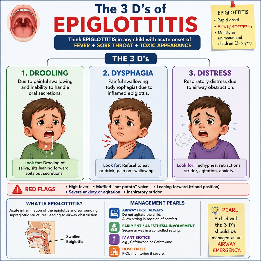
“3 D’s” of Epiglottitis

  1. Drooling
  2. Dysphagia
  3. Distress (respiratory)

Diagnosis

Clinical Diagnosis

  • Do not aggressively examine throat in severe cases because it may worsen airway obstruction.

Investigations

  • Laryngoscopy
  • Neck X-ray → Thumb sign
  • Blood culture/throat swab
  • Pulse oximetry (Mayo Clinic)

Management

Emergency Management

  1. Secure airway first
    • Oxygen
    • Endotracheal intubation if needed
    • Rarely tracheostomy
  2. Medications
    • IV antibiotics
    • Corticosteroids
    • IV fluids
  3. ICU monitoring

Complications

  • Sudden airway obstruction
  • Respiratory failure
  • Sepsis
  • Death if untreated (Mayo Clinic)

Prevention

  • Hib vaccination is the best preventive measure.
  • Good hygiene and infection control.

Difference Between Epiglottitis and Croup

FeatureEpiglottitisCroup
OnsetSuddenGradual
FeverHighMild
CoughUsually absentBarking cough
DroolingPresentRare
VoiceMuffledHoarse
AgeOlder children/adultsYoung children
EmergencySevere emergencyUsually mild

Key Points to Remember

  • Life-threatening airway emergency.
  • Drooling + stridor + muffled voice = suspect epiglottitis.
  • Do not force throat examination.
  • Airway management is priority.
  • Hib vaccine greatly reduced cases.

Epiglottitis

8 Clinical Features of Hypertrophic Pyloric Stenosis (HPS) — High-Yield Notes

Definition

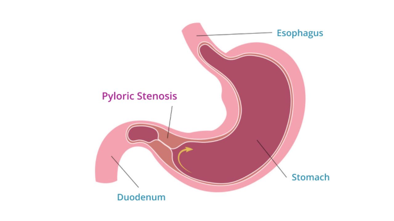
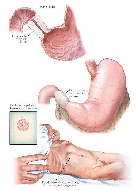
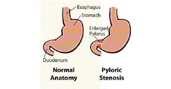
  • Hypertrophy + hyperplasia of pyloric muscle → gastric outlet obstruction in infants.

Epidemiology

  • Age: 2–8 weeks (classically 3–6 weeks)
  • More common in:
    • First-born males
    • Formula-fed infants
    • Positive family history
  • Associated with macrolide exposure (e.g., erythromycin)

Pathology

  • Thickened pyloric muscle narrows pyloric canal.
  • Causes obstruction to gastric emptying.

Clinical Features

Classic Triad

  1. Projectile non-bilious vomiting
  2. Visible gastric peristalsis
  3. Olive-shaped mass in epigastrium/right upper abdomen

Other Findings

  • Hungry after vomiting (“hungry vomiter”)
  • Weight loss/dehydration
  • Constipation
  • Failure to thrive

Why Vomiting is Non-Bilious?

  • Obstruction is proximal to duodenum → bile cannot enter vomitus.

Electrolyte Abnormality (Very Important)

Due to repeated vomiting:

  • Hypochloremic
  • Hypokalemic
  • Metabolic alkalosis

Mnemonic:

“Vomiting loses HCl”


Diagnosis

Investigation of Choice

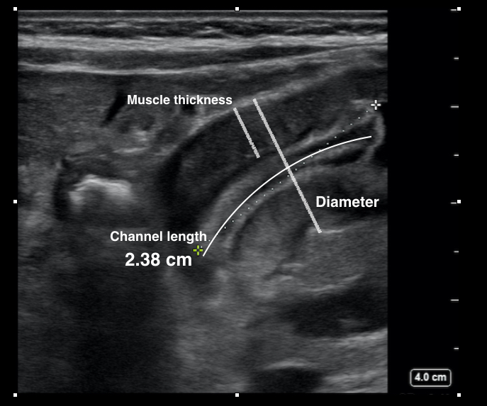
  • Ultrasound abdomen

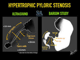
USG Findings

  • Thickened pylorus
  • Elongated pyloric canal

X-ray/Barium

  • “String sign” (narrowed pyloric canal)

Management

Initial

  • Correct:
    • Dehydration
    • Electrolyte imbalance

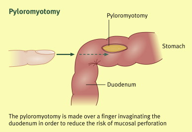
Definitive Treatment

  • Ramstedt pyloromyotomy

Important Differentials

  • Gastroesophageal reflux
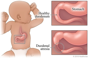
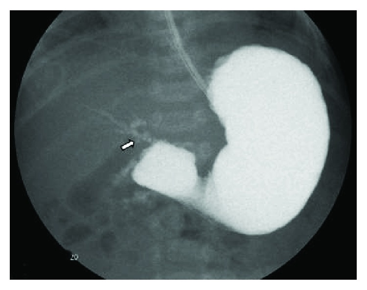
  • Duodenal atresia (bilious vomiting)
  • Intestinal obstruction
  • Sepsis/metabolic disorders

Super High-Yield One-Liners

  • Projectile non-bilious vomiting in a 3-week-old male = HPS until proven otherwise
  • Olive mass + visible peristalsis = classic clue
  • Best diagnostic test = Ultrasound
  • Treatment = Pyloromyotomy
  • Metabolic alkalosis is the classic acid-base disorder

Quick Revision Table

FeatureHPS
Age2–8 weeks
VomitingProjectile, non-bilious
AppetiteHungry after vomiting
MassOlive-shaped
DiagnosisUltrasound
ElectrolytesHypochloremic hypokalemic metabolic alkalosis
TreatmentRamstedt pyloromyotomy

Cesarean Section Delivery: Healing Time, 5 easy Recovery Tips, and Complication Prevention (cesarean section scar)

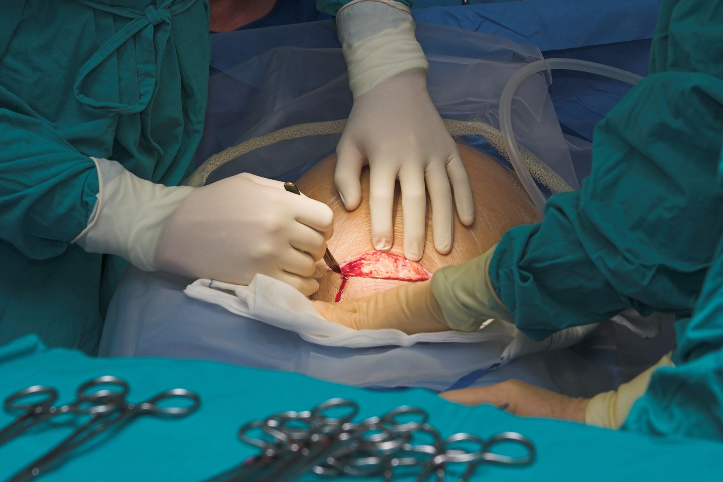
A cesarean section, commonly called a C-section, is a surgical procedure used to deliver a baby through incisions made in the mother’s abdomen and uterus. It may be planned in advance or performed as an emergency procedure when vaginal delivery could pose risks to the mother or baby.

C-sections are common worldwide and can be life-saving. However, because they involve major abdominal surgery, recovery usually takes longer than recovery after a vaginal birth. Understanding the healing process and following proper post-operative care can help mothers recover more comfortably and reduce the risk of complications.


What Happens During a Cesarean Section?

During a C-section, doctors make an incision through the abdominal wall and uterus to safely deliver the baby. The procedure is typically performed under spinal or epidural anesthesia, allowing the mother to remain awake while avoiding pain.

Common reasons for a cesarean delivery include:

  • Prolonged or difficult labor
  • Fetal distress
  • Multiple pregnancies (twins or more)
  • Breech position
  • Placenta-related complications
  • Previous cesarean delivery
  • Certain maternal health conditions

Healing Time After a C-Section

Recovery varies from person to person, but most women follow a general healing timeline.

First 24–48 Hours

  • Mothers are closely monitored in the hospital.
  • Pain, fatigue, and abdominal soreness are common.
  • Walking is encouraged within a day to improve circulation and prevent blood clots.

First 2 Weeks

  • Incision pain gradually decreases.
  • Light movement becomes easier.
  • Bleeding and discharge may continue.
  • Rest is essential.

4–6 Weeks

  • Most tissues heal significantly during this period.
  • Many women can return to light household activities.
  • Driving and moderate activity may resume after medical approval.

6–12 Weeks

  • Internal healing continues.
  • Energy levels improve.
  • Exercise can slowly restart with a doctor’s guidance.

Even after the external scar appears healed, internal tissues may still be recovering. Full recovery can sometimes take several months.


Common Symptoms During Recovery

The following symptoms are usually normal after a C-section:

  • Mild to moderate incision pain
  • Cramping
  • Vaginal bleeding
  • Fatigue
  • Swelling
  • Difficulty standing fully upright initially
  • Temporary numbness around the incision

However, worsening symptoms should never be ignored.


Tips for Faster Recovery

1. Get Adequate Rest

Sleep and rest are essential for tissue repair and hormonal recovery. New mothers should rest whenever the baby sleeps and avoid overexertion.

2. Walk Regularly

Gentle walking improves blood circulation, reduces gas pain, and lowers the risk of blood clots. Short walks several times daily are beneficial.

3. Support the Incision

Holding a pillow against the abdomen while coughing, laughing, or standing can reduce discomfort and protect the incision.

4. Stay Hydrated

Drinking enough water supports healing, digestion, and breast milk production.

5. Eat Nutritious Foods

A balanced diet rich in protein, iron, vitamins, and fiber helps tissue repair and prevents constipation.

Helpful foods include:

  • Lean meats and eggs
  • Fruits and vegetables
  • Whole grains
  • Yogurt
  • Nuts and seeds

6. Avoid Heavy Lifting

For at least 6 weeks, mothers should avoid lifting anything heavier than the baby.

7. Take Medications as Prescribed

Pain medicines and antibiotics should be taken exactly as directed.

8. Keep the Incision Clean and Dry

Gentle cleaning and proper drying reduce infection risk. Tight clothing should be avoided if it irritates the wound.

9. Accept Help From Others

Support from family members can reduce physical strain and emotional stress during recovery.

10. Attend Follow-Up Appointments

Regular medical checkups help ensure proper healing and early detection of complications.


Preventing Complications

While most women recover well, complications can occur if proper care is neglected.

Preventing Infection

Signs of infection include:

  • Redness
  • Swelling
  • Fever
  • Pus or foul-smelling discharge
  • Increasing pain

To prevent infection:

  • Wash hands before touching the incision.
  • Follow wound-care instructions carefully.
  • Avoid soaking in bathtubs until approved by a doctor.

Preventing Blood Clots

After surgery, blood clot risk increases temporarily.

Prevention measures include:

  • Early walking
  • Leg exercises
  • Staying hydrated
  • Wearing compression stockings if recommended

Preventing Constipation

Pain medications and reduced movement may slow digestion.

Helpful strategies:

  • Drink water
  • Eat fiber-rich foods
  • Walk regularly
  • Use stool softeners if prescribed

Emotional Health Matters

Some mothers experience anxiety, sadness, or emotional overwhelm after delivery.

Seek medical support if symptoms include:

  • Persistent sadness
  • Loss of interest
  • Severe mood swings
  • Difficulty bonding with the baby
  • Thoughts of self-harm

Postpartum depression is treatable and should never be ignored.


When to Seek Immediate Medical Care

A doctor should be contacted immediately if any of the following occur:

  • High fever
  • Heavy bleeding
  • Severe abdominal pain
  • Chest pain or breathing difficulty
  • Swelling or pain in one leg
  • Opening of the incision
  • Foul-smelling wound drainage
  • Persistent vomiting

These symptoms may indicate serious complications that require urgent treatment.


Long-Term Recovery and Scar Healing

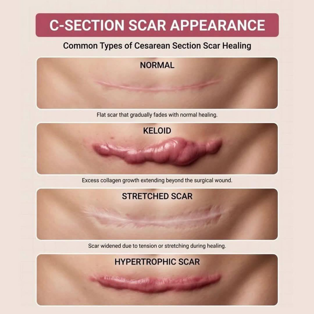
C-section scars usually fade gradually over time. Gentle scar care after complete wound closure may improve appearance.

Long-term recovery tips include:

  • Gradually rebuilding core strength
  • Maintaining a healthy weight
  • Avoiding smoking
  • Discussing future pregnancy plans with a healthcare provider

Many women go on to have healthy future pregnancies and deliveries after a cesarean section.


Conclusion

A cesarean section is a major surgical procedure that requires patience, rest, and proper care during recovery. Most mothers heal well within several weeks, especially when they follow healthy recovery habits and seek medical help promptly when needed.

Good nutrition, gentle movement, incision care, emotional support, and regular medical follow-up all play important roles in faster healing and complication prevention. With appropriate care and support, mothers can recover safely while focusing on bonding with their newborn and adjusting to life after childbirth.

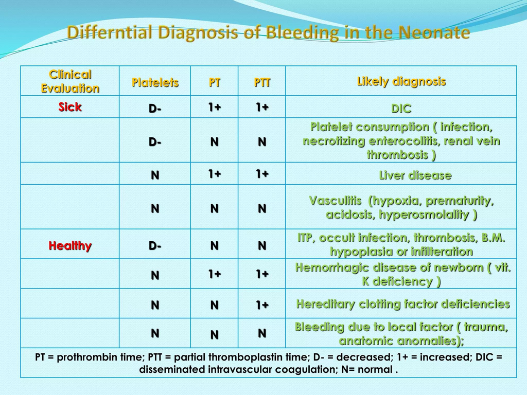
Bleeding and Increased PT/INR in Neonates: 10 Major Causes You Must Know

Prolonged PT/INR in neonates usually reflects abnormalities in the extrinsic and common coagulation pathways (Factors I, II, V, VII, X).
The causes can be grouped into physiologic, nutritional, hepatic, consumptive, inherited, and iatrogenic causes.


1. Physiological Causes

Physiologic immaturity of coagulation system

  • Normal neonates (especially preterms) have:
    • Lower vitamin K–dependent clotting factors
    • Reduced hepatic synthesis
  • Mild PT prolongation may occur in:
    • Prematurity
    • Very low birth weight infants

2. Vitamin K Deficiency (Most Important Cause)

Vitamin K Deficiency Bleeding (VKDB)

Most common clinically important cause of prolonged PT in neonates.

Why PT increases first?

Factor VII has the shortest half-life → PT prolongs earliest.

Risk factors

  • No vitamin K prophylaxis at birth
  • Exclusive breastfeeding
  • Maternal anticonvulsants:
    • Phenytoin
    • Phenobarbital
    • Carbamazepine
  • Maternal anti-TB drugs:
    • Rifampicin
    • Isoniazid
  • Malabsorption/cholestasis
  • Prolonged antibiotics

Types

TypeTiming
Early VKDB<24 hr
Classical VKDBDay 2–7
Late VKDB2 weeks–6 months

Labs

  • ↑ PT/INR (earliest finding)
  • Later ↑ aPTT
  • Normal platelets initially
  • Rapid correction after vitamin K

3. Liver Disease / Hepatic Dysfunction

Neonatal liver dysfunction reduces clotting factor synthesis.

Causes

  • Neonatal hepatitis
  • Biliary atresia
  • Cholestatic liver disease
  • Sepsis-associated liver dysfunction
  • Hypoxic ischemic injury
  • Metabolic liver disease
    • Galactosemia
    • Tyrosinemia

Labs

  • ↑ PT/INR
  • May not correct with vitamin K if severe hepatocellular failure

4. Disseminated Intravascular Coagulation (DIC)

Consumptive coagulopathy causing depletion of clotting factors.

Common neonatal triggers

  • Sepsis
  • Birth asphyxia
  • NEC
  • Shock
  • Severe hypoxia
  • Meconium aspiration
  • Severe hemolysis

Labs

  • ↑ PT
  • ↑ aPTT
  • ↓ fibrinogen
  • ↓ platelets
  • ↑ D-dimer/FDP

5. Sepsis

Can prolong PT due to:

  • Hepatic dysfunction
  • DIC
  • Cytokine-mediated coagulopathy
  • Vitamin K deficiency from poor feeding/antibiotics

6. Congenital Coagulation Factor Deficiencies

Factor VII deficiency

  • Isolated prolonged PT
  • Normal aPTT

Deficiencies affecting common pathway

  • Factor II deficiency
  • Factor V deficiency
  • Factor X deficiency
  • Fibrinogen disorders

Clues

  • Family history
  • Bleeding despite vitamin K
  • Persistent abnormal PT

7. Massive Bleeding / Dilutional Coagulopathy

Occurs after:

  • Massive transfusion
  • Exchange transfusion
  • Severe hemorrhage

Mechanism:

  • Dilution of clotting factors

8. Anticoagulant Exposure

Maternal drug exposure

  • Warfarin
  • Anticonvulsants
  • Anti-TB drugs

Neonatal medications

  • Heparin contamination of sample (usually affects aPTT more)

9. Cholestasis / Fat Malabsorption

Leads to poor vitamin K absorption.

Causes

  • Biliary atresia
  • Neonatal cholestasis
  • Cystic fibrosis
  • Intestinal disease

10. Laboratory / Sampling Errors

Always exclude before extensive workup.

Causes

  • Underfilled citrate tube
  • Clotted sample
  • Heparin contamination
  • Difficult sampling

Important High-Yield Pattern Recognition

PatternLikely Cause
Isolated ↑ PTVitamin K deficiency, Factor VII deficiency
↑ PT + ↑ aPTTLiver disease, DIC, severe VKDB
↑ PT + thrombocytopeniaDIC, sepsis
PT corrects after vitamin KVKDB
PT not correcting with vitamin KLiver failure or factor deficiency

High-Yield Neonatal Causes to Remember

  1. Vitamin K deficiency
  2. Sepsis
  3. DIC
  4. Liver disease/cholestasis
  5. Prematurity
  6. Congenital factor deficiency
  7. Maternal drug exposure

🚨 Increased PT/INR in Neonates — High Yield NICU Causes

🩸 Common causes of prolonged PT/INR in newborns:

▪️Vitamin K deficiency (most common)
▪️Sepsis
▪️DIC
▪️Liver disease / cholestasis
▪️Prematurity (physiologic immaturity)
▪️Congenital factor deficiencies (especially Factor VII)
▪️Maternal drugs
  • Anticonvulsants
  • Anti-TB drugs
  • Warfarin
▪️Massive bleeding / dilutional coagulopathy
▪️Laboratory sampling errors

⚡ High-yield tip:
➡️ PT rises EARLY in vitamin K deficiency because Factor VII has the shortest half-life.

🧠 Pattern to remember:
🔹 Isolated ↑ PT → Vitamin K deficiency / Factor VII deficiency
🔹 ↑ PT + ↑ aPTT → DIC or liver disease
🔹 Corrects after vitamin K → VKDB likely

PT/INR evaluates the extrinsic coagulation pathway and is commonly used in neonatal coagulation assessment. (bmcpediatr.biomedcentral.com)

6 Well Known Step Pathogenesis of Apnea of Prematurity Based on Nelson and Clohery to Clear Your Concept

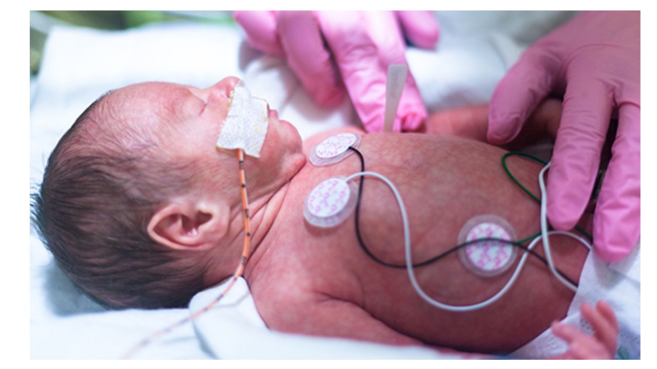
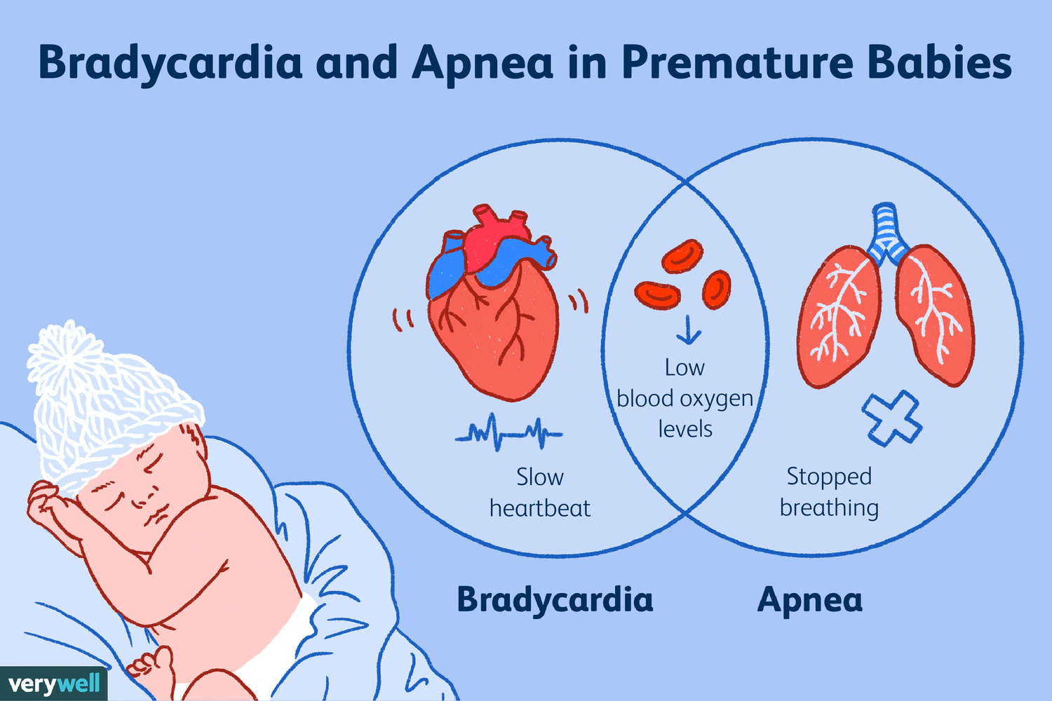
Apnea of prematurity (AOP) is a common condition where infants born before 37 weeks gestation experience breathing pauses lasting 15-20 seconds or more, often accompanied by slow heart rates (bradycardia) or low oxygen levels. It stems from an immature central nervous system and usually resolves on its own by 37–40 weeks postmenstrual age. Based on Cloherty and Stark’s Manual of Neonatal Care (9th Edition) and relevant pediatric algorithms and Nelson’s, here is a comprehensive summary of Apnea of Prematurity (AOP).

1. Definition and Classification

  • Definition: Apnea is the cessation of airflow. It is considered pathologic (an apneic spell) when:
    • Absence of airflow lasts 20 seconds or longer.
    • It is shorter than 20 seconds but accompanied by bradycardia (heart rate < 100 bpm) or hypoxemia (cyanosis or $SpO_2$ < 85–80%).
  • Classification:
    • Central: Total absence of inspiratory efforts (no diaphragmatic activity).
    • Obstructive: Inspiratory efforts persist, but airflow is blocked, usually at the pharyngeal level.
    • Mixed: A combination where airway obstruction precedes or follows central apnea. Most spells in preterm infants are mixed.

2. Incidence, Onset, and Duration

  • Incidence: Inversely related to gestational age (GA). It occurs in essentially all infants < 28 weeks’ GA and about 25% of those < 34 weeks’ GA.
  • Onset: Typically begins 1 to 2 days after birth. If spells do not occur within the first 7 days, AOP is unlikely to develop later unless triggered by other factors.
  • Duration: Usually ceases by 36 to 37 weeks’ postmenstrual age (PMA) in infants born at $\ge 28$ weeks, but frequently persists beyond term for those born more prematurely.

3. Pathogenesis (Underlying Mechanisms)

  • Developmental Immaturity: The primary cause is an immature central respiratory drive in the brainstem.
  • Sleep State: Spells are more frequent during active (REM) sleep, which is the predominant sleep state in preterms and is characterized by irregular breathing.
  • Chemoreceptor Response: Preterm infants have a decreased ventilatory response to increased $CO_2$ and may respond to hypoxia with hypoventilation rather than sustained hyperventilation.
  • Reflexes: Apnea can be triggered by stimulation of the posterior pharynx (e.g., vigorous suctioning), lung inflation, or fluid in the larynx.
  • Airway Mechanics: Poor muscle tone can lead to airway obstruction, especially during neck flexion or if there is nasal obstruction.
  • Note on GER: While gastroesophageal reflux is common in preterms, studies have not demonstrated an association between GER and AOP frequency.

4. Differential Diagnosis (Evaluation)

Apnea in a term infant or a “sick” preterm infant is always abnormal and requires looking for secondary causes:

  • Infection: Sepsis, meningitis, or necrotizing enterocolitis.
  • Metabolic Disorders: Hypoglycemia, hypocalcemia, or electrolyte imbalances (hyponatremia).
  • Neurologic: Intracranial hemorrhage (IVH), seizures, or birth asphyxia.
  • Impaired Oxygenation: PDA (Patent Ductus Arteriosus), anemia, or pneumonia.
  • Drugs: Maternal medications (magnesium, narcotics) or drug toxicity (e.g., phenobarbitone).

5. Management and Treatment

  • Monitoring: All infants < 35 weeks’ GA should be monitored for at least the first week. Monitor heart rate and $SpO_2$ in addition to respiration.
  • Immediate Action: Respond to the infant, not the monitor alarm. Most spells respond to tactile stimulation. If the infant is unresponsive, use bag-and-mask ventilation.
  • Positioning: Avoid extreme neck flexion or extension. Prone positioning may reduce apnea by stabilizing the chest wall.
  • Pharmacotherapy (Methylxanthines):
    • Caffeine Citrate: The drug of choice due to its long half-life (once-daily dosing), high therapeutic index, and lack of need for routine level monitoring.
    • Dosing: Loading dose of 20 mg/kg (10 mg/kg caffeine base), followed by a maintenance dose of 5–10 mg/kg daily.
    • Benefits: Reduces spells, the need for mechanical ventilation, and the risk of Bronchopulmonary Dysplasia (BPD).
  • Respiratory Support:
    • nCPAP (4–6 $cm H_2O$): Reduces mixed and obstructive spells by maintaining end-expiratory lung volume.
    • NIPPV: May be attempted if CPAP fails.
  • Other Considerations:
    • Blood Transfusion: May be considered if the hematocrit is < 25–30% and spells are frequent despite caffeine.
    • GER Treatment: Pharmacologic treatment of reflux (e.g., H2 blockers) is not recommended to treat AOP and may be harmful.

6. Discharge and Follow-up

  • Discharge Criteria: Infants should be free of significant apnea for 5 to 7 days after stopping caffeine.
  • Caffeine Offset: Because caffeine effects remain for up to a week, the “countdown” to discharge typically starts several days after the last dose.
  • Recurrence: Recurrent apnea can be triggered by viral illness, anesthesia, eye examinations, or immunizations. These infants should be monitored closely until at least 44 weeks’ PMA.
  • SIDS: A history of AOP does not increase the risk of Sudden Infant Death Syndrome (SIDS).