Absence of Anterior Fontanelle in a Newborn (Closed / Non-Palpable Anterior Fontanelle)

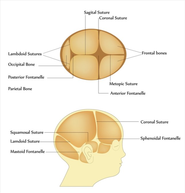
The anterior fontanelle is the largest fontanelle of the newborn skull and normally remains open during early infancy to allow brain growth and skull expansion.

1. Normal Anatomy and Physiology

  • The anterior fontanelle lies at the junction of:
    • Two frontal bones
    • Two parietal bones
  • Shape: Diamond-shaped
  • Average size at birth: 1–4 cm
  • Normal closure: 9–18 months

Functions:

  • Allows rapid brain growth
  • Facilitates molding during vaginal delivery
  • Serves as a clinical window for intracranial pressure assessment
normal fontanelle and suture in neonate

When the Anterior Fontanelle Is Absent at Birth

A non-palpable or absent anterior fontanelle suggests premature fusion of cranial sutures or abnormal skull ossification.

This finding must always be evaluated carefully because it may indicate craniosynostosis or underlying pathology.


Causes of Absent Anterior Fontanelle

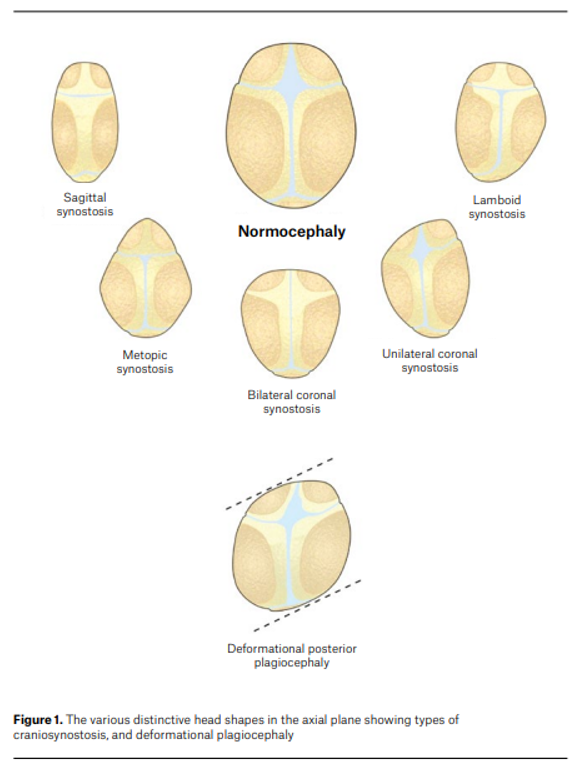
1. Craniosynostosis (Most Important Cause)

Premature fusion of one or more cranial sutures prevents normal skull expansion.

Types include:

Suture involvedResulting head shape
SagittalScaphocephaly (long narrow skull)
CoronalBrachycephaly
MetopicTrigonocephaly
Multiple suturesOxycephaly

Consequences:

  • Restricted skull growth
  • Raised intracranial pressure
  • Neurodevelopmental impairment if untreated

2. Hyperthyroidism (Congenital Thyrotoxicosis)

Seen in infants of mothers with Graves disease

Mechanism:

  • Increased thyroid hormone → accelerated bone maturation
  • Leads to early closure of sutures and fontanelles

Associated features:

  • Irritability
  • Tachycardia
  • Poor weight gain
  • Goiter
  • Exophthalmos (rare in neonates)

3. Microcephaly

Brain growth failure leads to small skull size, so sutures close early.

Common causes:

  • Intrauterine infections (TORCH)
  • Genetic syndromes
  • Severe hypoxic injury
  • Metabolic disorders

4. Skeletal Dysplasias

Some bone disorders cause abnormal skull ossification.

Examples:

  • Osteopetrosis
  • Thanatophoric dysplasia

5. Normal Variant

Rarely the fontanelle is very small or difficult to palpate, but sutures remain open and skull growth is normal.


Clinical Evaluation

1. History

Ask about:

Maternal history

  • Hyperthyroidism
  • Antithyroid drugs
  • TORCH infections

Perinatal history

  • Birth trauma
  • Neonatal illness

Family history

  • Craniosynostosis
  • Genetic syndromes

Developmental history

  • Feeding difficulty
  • Poor growth
  • Developmental delay

2. Physical Examination

Head Examination

Assess:

FeatureSignificance
Head circumferenceDetect microcephaly
Skull shapeSuggest specific craniosynostosis
Palpation of suturesCheck if fused or ridged
Remaining fontanellesPosterior fontanelle status

Look for Associated Signs

Neurologic:

  • Irritability
  • Vomiting
  • Bulging veins

Systemic:

  • Signs of hyperthyroidism
  • Dysmorphic features

Investigations

1. Imaging

Skull X-ray

  • Shows fused sutures

Cranial ultrasound

  • If some fontanelle is open

CT scan with 3D reconstruction

  • Gold standard for diagnosing craniosynostosis

2. Laboratory Tests

If systemic cause suspected:

TestPurpose
Thyroid function testDetect neonatal thyrotoxicosis
TORCH screeningIf infection suspected
Genetic testingSyndromic craniosynostosis

Complications

If due to craniosynostosis:

  • Raised intracranial pressure
  • Visual impairment
  • Developmental delay
  • Cognitive impairment

Management

1. Craniosynostosis

Referral to pediatric neurosurgery

Treatment:

  • Surgical cranial vault remodeling
  • Usually performed within first year of life

2. Neonatal Hyperthyroidism

Treat underlying condition:

  • Antithyroid drugs
  • Beta-blockers

3. Microcephaly

Management depends on cause:

  • Developmental support
  • Treat underlying infection/metabolic disease

Clinical Pearls (High-Yield)

  • Anterior fontanelle absent at birth → think craniosynostosis first.
  • Always measure head circumference.
  • Check skull shape and sutures carefully.
  • 3D CT scan confirms diagnosis.
  • Early surgical correction prevents intracranial hypertension and neurodevelopmental damage.

Creatine Kinase (CK) – High-Yield Medical Notes

1. Definition

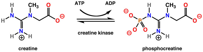
Creatine kinase (CK), also called creatine phosphokinase (CPK), is an intracellular enzyme that catalyzes the reversible conversion:Creatine+ATP↔Phosphocreatine+ADPCreatine + ATP \leftrightarrow Phosphocreatine + ADPCreatine+ATP↔Phosphocreatine+ADP

This reaction is crucial for energy storage and rapid regeneration of ATP, especially in muscle and brain tissues.


2. Location in Body

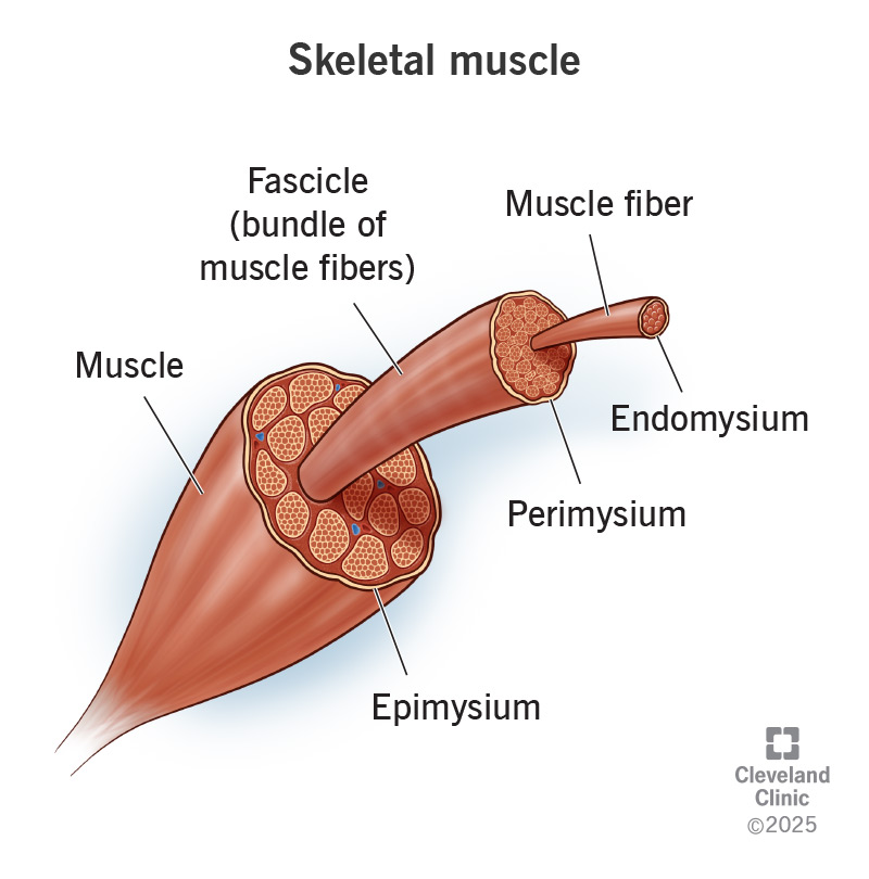
CK is present mainly in tissues with high energy demand:

TissueCK concentration
Skeletal muscleHighest
Cardiac muscleHigh
BrainModerate
Smooth muscleLow

Because CK is intracellular, serum CK rises when cell membrane damage occurs.


3. CK Isoenzymes

CK exists in three isoenzymes, formed by combinations of M (muscle) and B (brain) subunits.

IsoenzymeStructureMajor SourceClinical significance
CK-MMM + MSkeletal muscleMuscle injury, rhabdomyolysis
CK-MBM + BCardiac muscleMyocardial injury
CK-BBB + BBrain, smooth muscleCNS injury

Distribution

  • CK-MM: ~95–100% of total CK in normal serum
  • CK-MB: <5% normally
  • CK-BB: normally absent in serum

4. Normal Values

Normal ranges vary by lab.

Typical reference:

GroupCK (U/L)
Adult male50–200
Adult female40–150
ChildrenHigher than adults
NeonatesCan be very high after birth

5. Causes of Elevated CK

A. Skeletal Muscle Disorders

Most common cause.

Examples:

  • Muscular dystrophy (e.g., Duchenne muscular dystrophy)
  • Inflammatory myopathies
    • Polymyositis
    • Dermatomyositis
  • Trauma
  • Intramuscular injections
  • Vigorous exercise

B. Rhabdomyolysis

Massive CK elevation.

Causes:

  • Crush injury
  • Drugs (statins)
  • Heat stroke
  • Severe infections

CK may rise >5000–10,000 U/L.


C. Cardiac Causes

CK-MB rises in myocardial injury.

Example:

  • Myocardial Infarction

However, CK-MB is now largely replaced by troponins.


D. Neurologic Disorders

CK-BB may increase in:

  • Stroke
  • Brain trauma
  • Seizures

Example:

  • Stroke

E. Other Causes

  • Hypothyroidism
  • Alcohol abuse
  • Surgery
  • Prolonged immobilization
  • Severe infections

6. CK in Myocardial Infarction

Historically important marker.

ParameterCK-MB
Rise3–6 hours
Peak12–24 hours
Normalization48–72 hours

Used previously to detect reinfarction.

Now replaced mainly by troponin I/T.


7. CK in Pediatric Practice

Important uses:

Screening for muscular dystrophy

Very high CK in:

  • Duchenne muscular dystrophy
  • Becker muscular dystrophy

Levels may be 10–100× normal.


Evaluation of Hypotonia

Used when evaluating:

  • floppy infant
  • muscle weakness

Evaluation of Rhabdomyolysis

Symptoms:

  • myalgia
  • weakness
  • dark urine (myoglobinuria)

8. Causes of Decreased CK

Rare clinical significance.

Seen in:

  • Low muscle mass
  • Chronic illness
  • Pregnancy

9. Drugs Causing Elevated CK

Important clinically.

Examples:

DrugMechanism
StatinsMyopathy
AntipsychoticsNeuroleptic malignant syndrome
CorticosteroidsMuscle breakdown

Example condition:
Neuroleptic Malignant Syndrome


10. Investigation Panel When CK is Elevated

TestPurpose
CK-MBCardiac source
TroponinMI confirmation
AST / ALTMuscle vs liver
LDHTissue injury
Serum myoglobinRhabdomyolysis
Urine myoglobinKidney injury

11. CK in Rhabdomyolysis Severity

CK levelInterpretation
<1000Mild
1000–5000Moderate

5000 | Severe muscle injury |
15000 | High risk of renal failure |


12. Clinical Pearls (Exam High-Yield)

  • CK-MM → skeletal muscle injury
  • CK-MB → cardiac muscle
  • CK-BB → brain
  • Duchenne muscular dystrophy → CK extremely high
  • CK rises after muscle trauma/exercise
  • CK-MB replaced by troponin in MI diagnosis

✅ One-line exam memory tip

“CK rises with muscle damage — skeletal (MM), cardiac (MB), brain (BB).”

Complicated Vs Uncomplicated UTI (Notes) – Clinical Practice and Diagnosis


1. Definition

TypeDefinition
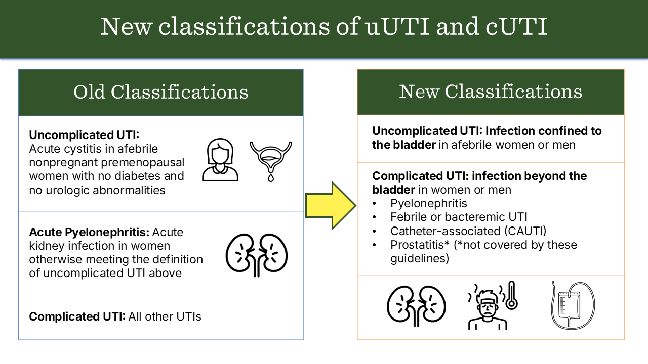
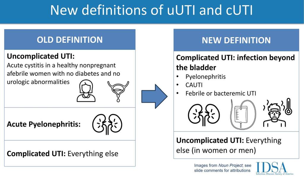
Uncomplicated UTIInfection of the lower urinary tract (bladder and urethra) occurring in an otherwise healthy, non-pregnant woman with a structurally and functionally normal urinary tract.
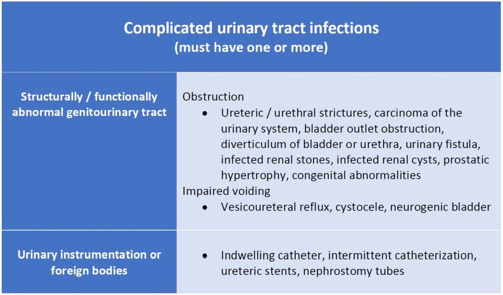
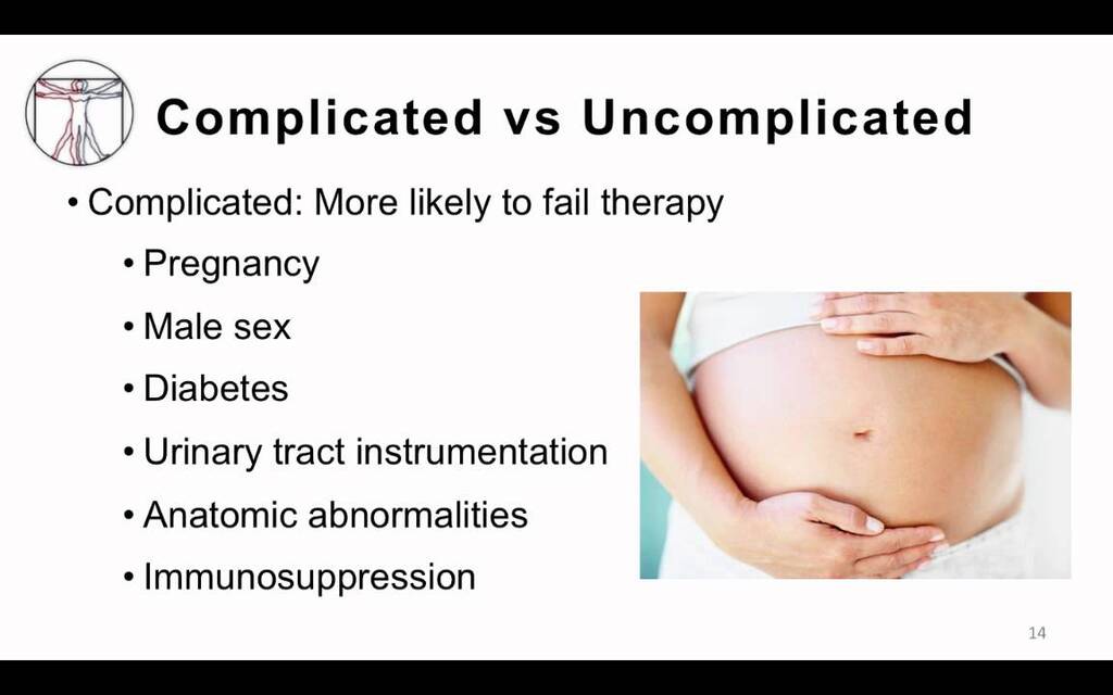
Complicated UTIUTI occurring in individuals with structural or functional abnormalities of the urinary tract, comorbidities, or other risk factors that increase risk of treatment failure or recurrence.

2. Typical Patients

TypeCommon in
UncomplicatedHealthy, premenopausal, non-pregnant females
ComplicatedMales, pregnant females, children, elderly, diabetics, catheterized patients, immunocompromised patients

3. Etiology

TypeCommon PathogensSpecial Pathogens
UncomplicatedE. coli (≈80–90%), Staphylococcus saprophyticusRarely Klebsiella, Proteus
ComplicatedE. coli, Klebsiella, Proteus, Enterobacter, Pseudomonas, Enterococcus, CandidaOften multidrug-resistant (MDR) organisms

4. Predisposing / Risk Factors

Uncomplicated:

  • Female gender (short urethra)
  • Sexual activity
  • Spermicides

Complicated:

  • Structural: Stones, strictures, obstruction (BPH, vesicoureteral reflux)
  • Functional: Neurogenic bladder, indwelling catheter
  • Systemic: Diabetes mellitus, immunosuppression
  • Male gender
  • Pregnancy

5. Clinical Presentation

TypeFeatures
UncomplicatedDysuria, frequency, urgency, suprapubic pain, no systemic signs
ComplicatedMay have fever, chills, flank pain, systemic toxicity, sepsis, poor response to therapy

6. Investigations

TypeLab Approach
UncomplicatedUrinalysis ± urine culture (often empirical treatment)
ComplicatedUrine culture mandatory, imaging (USG, CT KUB) if obstruction suspected, blood cultures if febrile

7. Treatment

TypeApproach
UncomplicatedShort-course oral antibiotics (3–5 days): Nitrofurantoin, TMP-SMX, Fosfomycin, Pivmecillinam
ComplicatedLonger course (7–14 days), guided by culture; IV therapy if severe (Ceftriaxone, Piperacillin-tazobactam, Carbapenems for MDR); treat underlying cause (remove catheter, relieve obstruction)

8. Prognosis

TypePrognosis
UncomplicatedExcellent, low recurrence with proper hygiene
ComplicatedRisk of recurrence, sepsis, renal scarring, abscess

9. Example Cases

ScenarioType
25-year-old woman with dysuria, no comorbiditiesUncomplicated
60-year-old diabetic man with fever, flank painComplicated
Pregnant woman with bacteriuriaComplicated
Patient with indwelling Foley catheter and feverComplicated

Summary Table

FeatureUncomplicatedComplicated
HostHealthy femaleAny comorbidity or abnormality
SiteLower UTI (cystitis)Any (cystitis, pyelonephritis, sepsis)
OrganismsUsually E. coliPolymicrobial, resistant organisms
TherapyShort courseLong course, guided by culture
PrognosisExcellentVariable, risk of recurrence

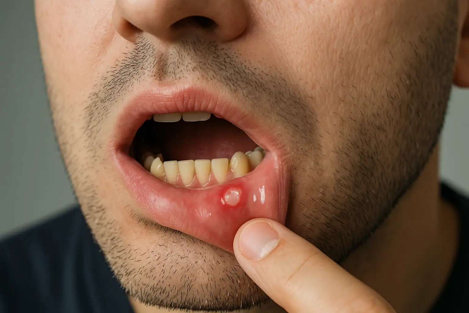
Aphthous Ulcers: Step-by-Step Workup and Management

1. Clinical Assessment

History

  • Onset, duration, frequency, and number of ulcers
  • Pain severity, triggers (trauma, stress, certain foods)
  • Systemic symptoms: fever, diarrhea, joint pain, fatigue
  • Family history of similar lesions
  • Medications (NSAIDs, beta-blockers)
  • Previous treatments and response

Examination

  • Location: non-keratinized mucosa (buccal, labial, ventral tongue, floor of mouth)
  • Size:
    • Minor (<1 cm) – most common
    • Major (>1 cm, deeper, may scar)
    • Herpetiform (multiple small ulcers)
  • Number: single or multiple
  • Appearance: round/oval, yellow-white pseudomembrane with erythematous halo
  • Rule out: secondary causes (herpes simplex, trauma, systemic disease)

2. Investigations (If atypical or recurrent)

  • Basic labs: CBC, iron studies, vitamin B12, folate (nutritional deficiencies)
  • CRP/ESR: if systemic symptoms
  • Serology: celiac disease (tTG-IgA), HIV (if risk factors)
  • Biopsy: rare, only if persistent >3 weeks, atypical, or suspected malignancy
  • Microbiology: rarely needed; ulcers are typically non-infectious

3. Classification

TypeFeaturesManagement Focus
Minor<1 cm, 1–5 lesions, heal 7–14 daysSymptomatic relief
Major>1 cm, deep, persistent, may scarSystemic therapy possible
HerpetiformMultiple, small, coalescing, painfulCombination topical/systemic therapy

4. Management

A. General Measures

  • Avoid triggers: spicy/acidic foods, trauma
  • Good oral hygiene
  • Avoid sodium lauryl sulfate in toothpaste
  • Stress management
  • Nutritional supplementation if deficient

B. Topical Therapy (First-line for minor ulcers)

  • Topical corticosteroids:
    • Triamcinolone acetonide 0.1% in orabase
    • Fluocinonide 0.05% gel
  • Topical anesthetics:
    • Lidocaine 2% gel for pain relief
  • Protective agents:
    • Orabase, hydroxypropyl cellulose films

C. Systemic Therapy (For severe, recurrent, or major ulcers)

  • Oral corticosteroids:
    • Prednisone short course (e.g., 30–60 mg/day tapering)
  • Immunomodulators (refractory cases):
    • Colchicine, thalidomide, dapsone
  • Address underlying systemic disease:
    • Nutritional deficiencies, IBD, Behçet’s disease, etc.

D. Adjunctive Therapy

  • Antimicrobial mouth rinses: chlorhexidine gluconate
  • Pain control: NSAIDs (if not contraindicated)
  • Laser therapy (experimental for pain reduction and healing)

5. Follow-Up

  • Minor ulcers: usually self-limiting; review if >3 weeks
  • Recurrent or major ulcers: monitor frequency, severity, and response to therapy
  • Evaluate for systemic disease if persistent or atypical

Approach to a Child with Wheeze

1. Definition

  • Wheeze is a high-pitched, musical, continuous sound produced by airflow through narrowed airways, typically during expiration.
  • Indicates airway obstruction at any level (from trachea to small bronchi).

2. Initial Assessment

A. ABC Assessment

  • Airway: Ensure patency; look for obstruction (secretions, foreign body).
  • Breathing:
    • Respiratory rate, effort (retractions, nasal flaring, grunting)
    • Oxygen saturation (SpO₂)
    • Presence and distribution of wheeze
    • Work of breathing (mild / moderate / severe)
  • Circulation: Heart rate, capillary refill time, BP, color.

B. Severity Assessment

SeverityFeatures
MildSpeaking in sentences, SpO₂ > 94%, mild wheeze
ModerateBreathless, feeding difficulty, SpO₂ 90–94%
SevereCannot talk/feed, SpO₂ < 90%, exhaustion, cyanosis, silent chest

3. Focused History

AspectKey Points
Onset & CourseSudden (foreign body) vs gradual (infection/asthma)
Frequency/PatternRecurrent vs first episode
TriggersViral infection, allergen, exercise, cold air, smoke
Associated SymptomsFever, cough, coryza, vomiting, feeding difficulty
Past Medical HistoryPrevious wheezing, atopy, eczema, prematurity, GERD
Family/Social HistoryAsthma, allergies, smoking, housing
Drug historyRecent medications, response to bronchodilators

4. Physical Examination

  • General: Distress, cyanosis, growth, hydration.
  • Respiratory system:
    • Chest expansion, use of accessory muscles
    • Percussion (hyperresonant / dull)
    • Auscultation:
      • Wheeze: polyphonic (asthma/viral), monophonic (focal obstruction)
      • Air entry: symmetrical or reduced
      • Crackles: suggest infection or bronchiolitis
  • Cardiac: Murmurs (congenital lesions)
  • Other systems: Skin (eczema), ENT (allergic rhinitis), clubbing.

5. Differential Diagnosis

Age GroupCommon Causes
Infant (<1 yr)Bronchiolitis, congenital airway anomalies, aspiration, GERD
Toddler (1–5 yr)Viral-induced wheeze, foreign body aspiration, asthma
Older child (>5 yr)Asthma, allergic bronchitis, chronic suppurative lung disease

6. Investigations

(Usually guided by clinical picture; many cases diagnosed clinically)

  • Pulse oximetry – essential.
  • Chest X-ray – if first episode, focal findings, suspicion of foreign body/pneumonia.
  • Blood tests – if severe or infection suspected.
  • Allergy testing / IgE – in recurrent or atopic cases.
  • Spirometry / Peak Flow – for older cooperative children (asthma diagnosis).
  • Bronchoscopy – if persistent localized wheeze or suspicion of foreign body.

7. Management

A. Immediate Management

  • Mild/Moderate:
    • Nebulized or inhaled salbutamol (via spacer or nebulizer)
    • Oxygen if SpO₂ < 94%
    • Oral prednisolone if known asthma or recurrent wheeze
  • Severe / Life-threatening:
    • High-flow oxygen
    • Nebulized salbutamol ± ipratropium bromide
    • IV steroids (hydrocortisone)
    • IV magnesium sulfate / aminophylline / salbutamol if poor response
    • Consider PICU referral

B. Underlying Cause

  • Bronchiolitis – supportive (O₂, fluids, suction)
  • Foreign body – urgent ENT/pulmonary referral
  • Asthma – follow stepwise management (as per BTS/SIGN or GINA)

8. Long-term Management

  • Identify triggers and educate parents on avoidance.
  • Asthma education: inhaler technique, action plan.
  • Follow-up to reassess control and adjust therapy.

9. Red Flags

  • Silent chest, exhaustion, cyanosis
  • Poor air entry or asymmetry
  • Persistent localized wheeze
  • Failure to thrive or recurrent pneumonia
  • Sudden onset without infection (→ foreign body)

10. Summary Table

StepKey Action
1ABC and severity assessment
2Focused history
3Physical examination
4Consider differential diagnoses
5Targeted investigations
6Manage acutely + treat cause
7Educate and follow-up

“The Mysterious Nosebleed Every Month: A Story of Vicarious Menstruation”


Background

It was a quiet morning at the outpatient clinic when 23-year-old Sita walked in with a puzzled expression. She looked perfectly healthy—until she spoke.

“Doctor,” she began, “every month when my friends get their periods, I don’t bleed like them. Instead, blood comes out from my nose.”

At first, everyone thought it was a coincidence. Maybe sinus problems? Maybe dry air? But the timing was too perfect—every 28 days, her nose would bleed for two or three days, right when her lower abdomen cramped and her mood shifted.

The Curious Case

After a few cycles of this strange pattern, Sita’s physician grew suspicious and ordered tests. Hormones were normal. Sinus scan—clean. Then came the question that changed everything:

“Does your nosebleed coincide with your menstrual cycle?”

When Sita nodded, the puzzle pieces clicked together. The diagnosis: Vicarious Menstruation—a rare and fascinating medical phenomenon where menstrual bleeding occurs from sites outside the uterus.

What Is Vicarious Menstruation?

Vicarious menstruation happens when the body’s endometrial-like tissue or hormone-sensitive mucosa outside the uterus responds to the same hormonal cycle that causes normal menstruation.

In simpler terms, when the uterine lining sheds, some other part of the body “joins in.”

Reported sites include:

  • Nose (most common – causing cyclical nosebleeds)
  • Lungs (causing hemoptysis or blood in sputum)
  • Skin (cyclical bruises or bleeding spots)
  • Eyes, ears, or even gastrointestinal tract in rare cases

How Does It Happen?

The exact cause isn’t fully understood, but doctors believe it may occur due to:

  • Hormonal sensitivity: Mucous membranes in certain organs respond to estrogen and progesterone fluctuations.
  • Endometrial implantation: Rarely, endometrial cells may reach other body sites via blood or lymphatic spread (similar to endometriosis).
  • Vascular fragility: Hormonal changes may increase capillary fragility during menses.

Sita’s Diagnosis and Treatment

Sita’s case was confirmed after observing the pattern for several months. ENT evaluation found fragile nasal capillaries that responded to hormone fluctuations.

Treatment involved hormonal regulation using oral contraceptive pills to suppress ovulation and stabilize estrogen levels. Within two months, her nosebleeds stopped—and normal menstruation returned.

Why It Matters

Vicarious menstruation reminds us that the human body is deeply interconnected. Hormones don’t just act in one place—they ripple through every organ, tissue, and mood.

For healthcare students and young clinicians, Sita’s story is a gentle warning:

“Always listen to the cycle. Not every period happens through the uterus.”

Takeaway for Readers

If someone experiences cyclical bleeding from any unusual site—nose, eyes, skin—it’s worth noting the timing. Keeping a menstrual diary can help doctors detect such patterns.

It’s rare, yes—but as Sita’s story shows, even the body’s most mysterious messages make sense when we listen carefully.