Diamond-Blackfan ane

Macrocytic Anemia: A Clinical Overview (Based on Nelson Textbook of Pediatrics)

Macrocytic anemia is a hematological condition characterized by the presence of enlarged red blood cells (RBCs), typically reflected by an increased mean corpuscular volume (MCV >100 fL). In pediatric practice, as outlined in Nelson Textbook of Pediatrics, macrocytic anemia represents an important diagnostic category that often signals underlying nutritional deficiencies, bone marrow disorders, or systemic disease.

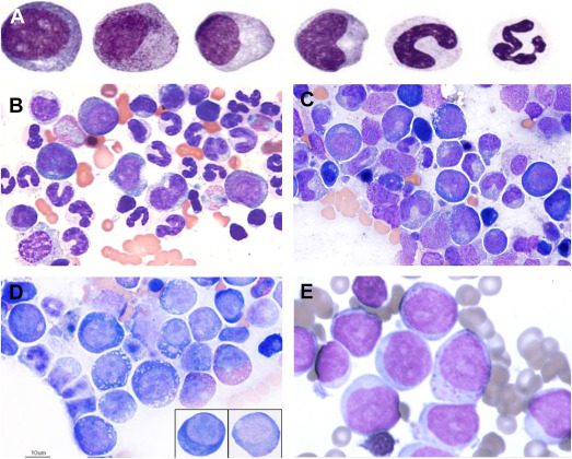
Classification of Macrocytic Anemia

Macrocytic anemia can broadly be divided into:

  1. Megaloblastic Macrocytic Anemia
    • Caused by impaired DNA synthesis
    • Characterized by megaloblasts in the bone marrow
  2. Non-Megaloblastic Macrocytic Anemia
    • Not associated with defective DNA synthesis
    • Often due to membrane abnormalities or increased reticulocyte count

Etiology

1. Megaloblastic Causes

  • Vitamin B12 deficiency
    • Dietary deficiency (rare in children unless strict vegan diet)
    • Malabsorption (e.g., pernicious anemia, ileal disease)
  • Folate deficiency
    • Poor dietary intake
    • Increased requirement (e.g., infancy, hemolytic anemia)
    • Malabsorption syndromes

2. Non-Megaloblastic Causes

  • Liver disease
  • Hypothyroidism
  • Reticulocytosis (e.g., hemolysis, blood loss)
  • Bone marrow disorders (e.g., aplastic anemia, myelodysplasia)
  • Drug-induced (e.g., anticonvulsants, chemotherapy)

Pathophysiology

In megaloblastic anemia, impaired DNA synthesis leads to delayed nuclear maturation while cytoplasmic development proceeds normally. This nuclear-cytoplasmic asynchrony results in large, immature erythroid precursors (megaloblasts) and macrocytic RBCs in circulation.

Vitamin B12 and folate are essential cofactors in DNA synthesis. Deficiency of either disrupts thymidine production, impairing cell division.


Clinical Features

Clinical presentation varies depending on severity and cause:

  • General symptoms
    • Pallor
    • Fatigue
    • weakness
  • Gastrointestinal
    • Glossitis (smooth, red tongue)
    • Poor appetite
  • Neurological (especially in B12 deficiency)
    • Paresthesia
    • Ataxia
    • Developmental delay (in infants)

Laboratory Findings

  • Elevated MCV (>100 fL)
  • Hypersegmented neutrophils (hallmark of megaloblastic anemia)
  • Low hemoglobin
  • Peripheral smear: macro-ovalocytes
  • Serum levels:
    • ↓ Vitamin B12
    • ↓ Folate
  • Elevated homocysteine (both deficiencies)
  • Elevated methylmalonic acid (specific for B12 deficiency)

Diagnosis

Diagnosis involves:

  1. Complete blood count (CBC)
  2. Peripheral blood smear
  3. Serum vitamin B12 and folate levels
  4. Bone marrow examination (if unclear or severe cases)

Management

Vitamin B12 Deficiency

  • Parenteral B12 (cyanocobalamin or hydroxocobalamin)
  • Lifelong therapy in irreversible causes

Folate Deficiency

  • Oral folic acid supplementation
  • Treat underlying cause

⚠️ Important: Always rule out B12 deficiency before treating folate deficiency alone, as folate can worsen neurological symptoms of B12 deficiency.


Complications

  • Neurological deficits (irreversible if untreated)
  • Growth retardation in children
  • Increased risk of infections due to pancytopenia

Prevention

  • Adequate dietary intake (green leafy vegetables, animal products)
  • Supplementation in high-risk groups
  • Early detection in infants and children

Conclusion

Macrocytic anemia in children requires a systematic approach to identify underlying causes. As emphasized in Nelson Textbook of Pediatrics, distinguishing between megaloblastic and non-megaloblastic anemia is critical for appropriate management. Early diagnosis and treatment can prevent serious complications, particularly irreversible neurological damage in vitamin B12 deficiency.

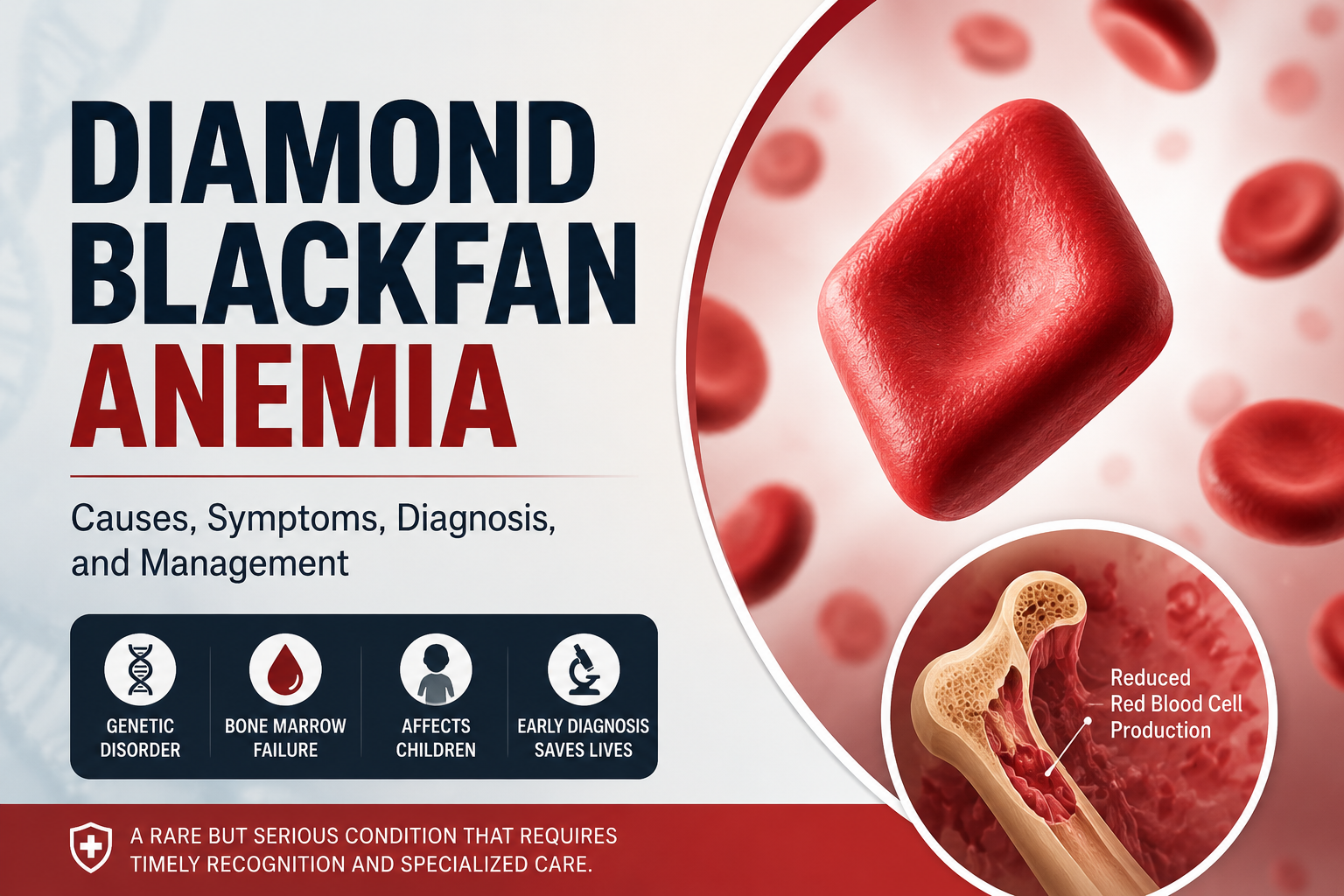
Diamond-Blackfan Anemia 101: Everything You Need to Know

Diamond-Blackfan anemia (DBA) is a rare congenital blood disorder characterized by the failure of the bone marrow to produce red blood cells. It usually presents in infancy and is classified as a congenital pure red cell aplasia. DBA is notable for its genetic basis, variable physical malformations, and lifelong management challenges.

Key facts

  • Onset: Typically within the first year of life
  • Genetic cause: Mutations in ribosomal protein genes
  • Inheritance pattern: Autosomal dominant (most cases de novo)
  • Prevalence: About 5–7 per million live births
  • Treatment options: Corticosteroids, chronic transfusions, or stem cell transplantation

Pathophysiology

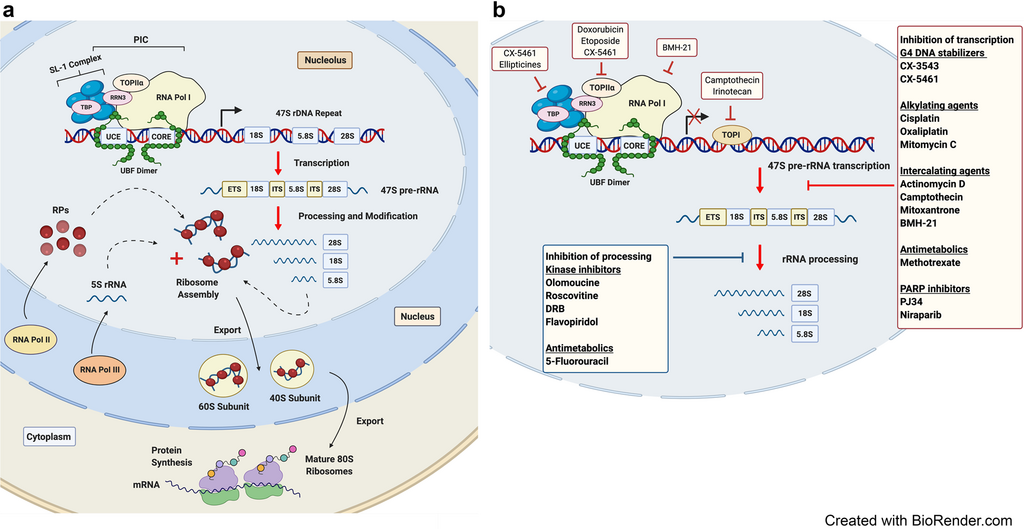
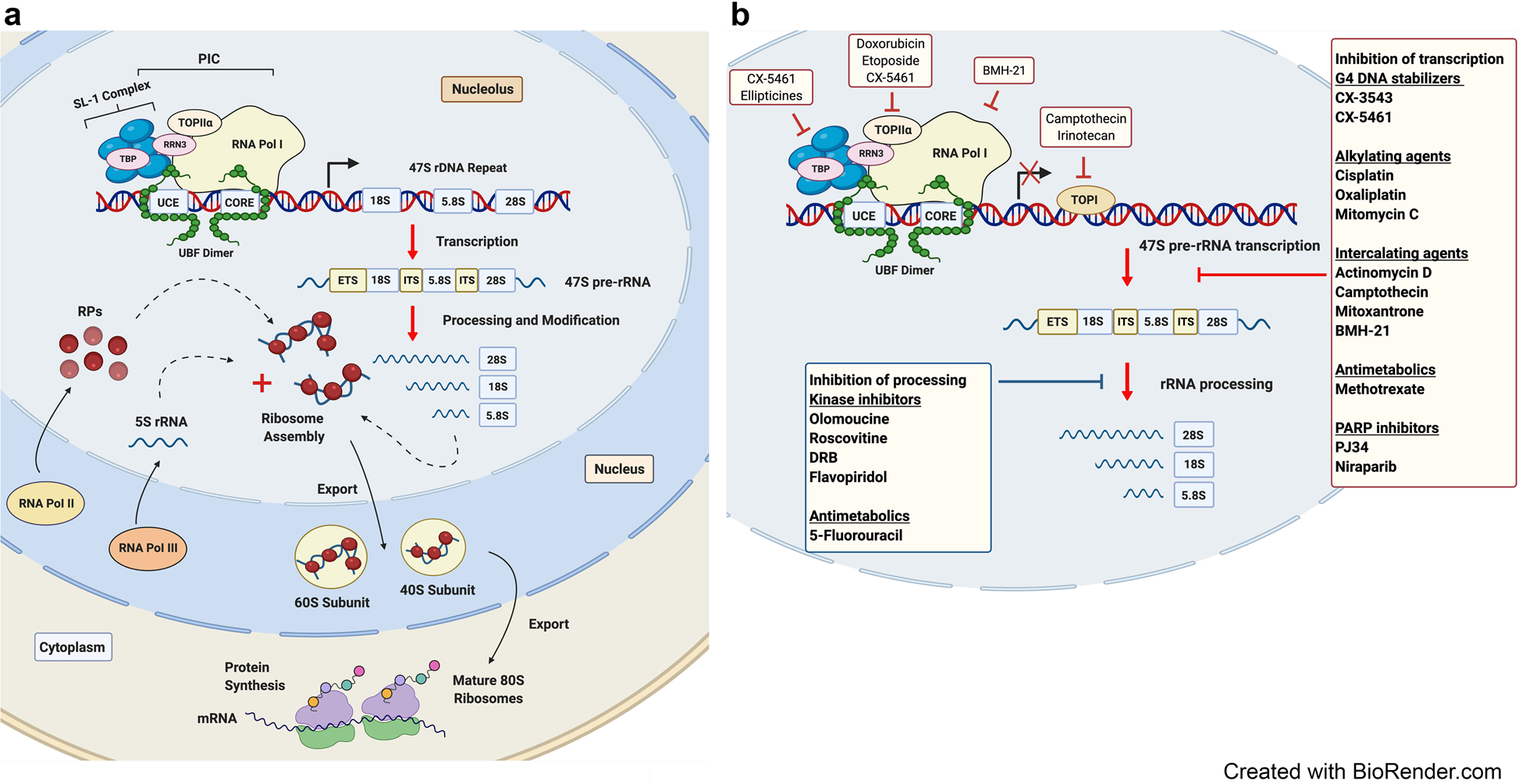
Diamond-Blackfan anemia arises from mutations that impair ribosome biogenesis, leading to defective erythroid progenitor development. The bone marrow becomes selectively deficient in red cell precursors, while white cells and platelets remain normal. Most cases involve mutations in genes encoding ribosomal proteins such as RPS19, RPL5, or RPL11, disrupting protein synthesis and cellular growth.

Clinical features

Infants with DBA commonly present with pallor and anemia. Physical anomalies are present in about half of cases, including craniofacial abnormalities, thumb or limb malformations, and heart or kidney defects. Growth retardation and an increased lifetime risk of malignancies such as leukemia and osteogenic sarcoma are recognized complications.

8 Vitamins that men with weak erections should take

Diagnosis

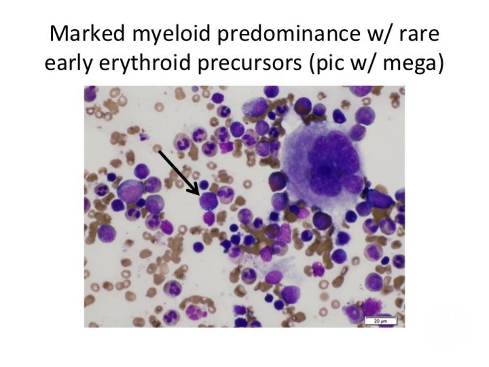
Diagnosis combines hematologic findings—macrocytic anemia, reticulocytopenia, and normal marrow cellularity except for absent red cell precursors—with genetic testing for ribosomal protein gene mutations. Elevated erythrocyte adenosine deaminase (eADA) activity is a common biomarker.

Management and prognosis

Initial treatment often involves corticosteroids to stimulate red cell production. Patients unresponsive to steroids may require regular transfusions with iron chelation therapy to prevent overload, or hematopoietic stem cell transplantation as a potential cure. Advances in genetic understanding have improved prognosis, but lifelong monitoring remains essential due to treatment complications and cancer risk.

Thyroid hormones, World Thyroid day 2023

Does Sex Increase Breast or Butt Size in Women?

(A Scientific, Myth-Busting Blog with Visual Explanation)


Introduction

This is one of the most common myths people believe:
👉 “Regular sex makes breasts and butt bigger.”

It sounds believable—because sex involves hormones, body changes, and physical stimulation. But what does actual medical science say?

Let’s break it down with evidence.


What Happens to the Body During Sex?

7

During sexual arousal and orgasm, several temporary physiological changes occur:

Increased blood flow

  • Blood vessels dilate (vasocongestion)
  • Breasts may appear fuller and slightly enlarged

Hormonal release

  • Oxytocin, dopamine, estrogen, progesterone rise
  • Nipples become erect and more sensitive

Temporary swelling effect

  • Breasts can appear 15–25% larger during arousal

👉 But here’s the key:
These changes are temporary and reverse after sex.


Does Sex Cause Permanent Breast Growth?

Short answer: No.

Scientific evidence clearly shows:

  • Breast size is mainly determined by:
    • Genetics
    • Body fat
    • Hormonal changes (puberty, pregnancy, medications)
  • Sex does NOT cause permanent breast enlargement

Even repeated sexual activity does not stimulate long-term tissue growth.


What Actually Increases Breast Size?

6

Real, evidence-based factors:

1. Genetics

  • The biggest determinant of size and shape

2. Body weight / fat

  • Breasts are largely fatty tissue
  • Weight gain → size increase

3. Hormones

  • Estrogen & progesterone drive growth
  • Seen during:
    • Puberty
    • Pregnancy
    • Hormonal therapy

4. Pregnancy & lactation

  • Causes true enlargement due to glandular growth

What About Buttocks Size?

6

Does sex increase butt size?

No — not directly.

Buttocks size depends on:

  • Fat distribution (influenced by estrogen)
  • Muscle mass (gluteal muscles)
  • Genetics
  • Exercise (e.g., squats, hip thrusts)

Sex does not stimulate fat deposition or muscle growth in the buttocks.

Why Do People Believe This Myth?

Several reasons:

1. Temporary swelling during sex

  • Breasts look bigger → mistaken as permanent

2. Hormonal timing confusion

  • Women may notice changes during:
    • Ovulation
    • Menstrual cycle
  • These are hormone-driven, not sex-driven

3. Cultural myths

  • Similar to:
    • “Marriage increases breast size” (also false)

Final Scientific Verdict

What sex DOES do:

  • Temporary breast fullness
  • Nipple erection
  • Increased sensitivity
  • Short-term hormonal fluctuations

What sex DOES NOT do:

  • Permanently increase breast size
  • Increase butt size
  • Change body fat distribution

Clinical Bottom Line (For Medical Perspective)

  • Breast size = fat + glandular tissue + hormones
  • Butt size = fat distribution + muscle mass
  • Sex = transient physiological response only

Conclusion

Sex may make breasts look bigger for a short time, but it does not cause real growth.
Butt size is completely unrelated to sexual activity.

This is a classic biological myth—explained by temporary changes and misunderstood hormones.

The Complete Clinical Approach to Vomiting in Pediatrics

A structured, practical guide for clinicians


Introduction

Vomiting is one of the most common presenting complaints in pediatric practice—ranging from benign, self-limiting illnesses to life-threatening surgical and metabolic emergencies. It is not a diagnosis, but a symptom with a broad differential, involving gastrointestinal (GI), neurological, metabolic, infectious, and psychological causes.

The real challenge is not treating vomiting—but identifying which child is sick and why.


Understanding Vomiting

Vomiting is a protective reflex involving coordinated contraction of abdominal muscles and relaxation of the lower esophageal sphincter, leading to expulsion of gastric contents.

Types of Vomiting

  • Acute vomiting → hours to days (e.g., gastroenteritis)
  • Chronic vomiting → weeks (e.g., GERD, metabolic causes)
  • Cyclic vomiting → episodic with symptom-free intervals
  • Projectile vomiting → suggests obstruction (e.g., pyloric stenosis)
  • Bilious vomiting (green) → surgical emergency until proven otherwise

Step 1: Initial Stabilization (ABC First)

Before thinking of diagnosis:

  • Airway → risk of aspiration
  • Breathing → respiratory distress?
  • Circulation → shock, dehydration

Assess:

  • Vitals
  • Capillary refill
  • Level of consciousness
  • Hydration status

👉 This step is critical because vomiting can rapidly lead to dehydration and electrolyte imbalance.


Step 2: Identify RED FLAG Signs

These determine urgency and need for immediate intervention:

Major Red Flags

  • Bilious (green) vomiting
  • Bloody vomiting
  • Altered sensorium
  • Severe dehydration
  • Persistent projectile vomiting
  • Abdominal distension or peritonitis
  • Inconsolable crying (infants)
  • Neck stiffness + fever (meningitis)
  • Morning vomiting + headache (raised ICP)

These features suggest serious pathology such as:

  • Intestinal obstruction
  • Intussusception
  • Meningitis
  • Intracranial hypertension
  • Appendicitis

Step 3: Age-Based Differential Diagnosis

Age is one of the most powerful diagnostic clues.

1. Neonates (0–28 days)

Think danger first:

  • Intestinal obstruction (atresia, malrotation)
  • Hirschsprung disease
  • Sepsis
  • Inborn errors of metabolism

👉 Bilious vomiting = surgical emergency


2. Infants

  • Gastroesophageal reflux (common)
  • Pyloric stenosis → projectile vomiting
  • Intussusception
  • Food allergy
  • Infection

3. Children

  • Acute gastroenteritis (most common)
  • Appendicitis
  • UTI
  • Pneumonia (post-tussive vomiting)
  • Migraine

4. Adolescents

  • Pregnancy
  • Eating disorders
  • Drug/toxin ingestion
  • Diabetic ketoacidosis (DKA)
  • Intracranial causes

Step 4: Focused History

A good history often gives the diagnosis.

Key Questions

  1. Onset
    • Sudden → infection, obstruction
    • Chronic → GERD, metabolic
  2. Character of Vomit
    • Bilious → obstruction
    • Projectile → pyloric stenosis
    • Blood → gastritis, ulcer
  3. Relation to Feeding
    • Immediately after feeds → reflux
    • Delayed → obstruction
  4. Associated Symptoms
    • Fever → infection
    • Diarrhea → gastroenteritis
    • Headache → intracranial cause
    • Abdominal pain → surgical cause
  5. Systemic Clues
    • Poor weight gain → chronic disease
    • Polyuria → DKA
    • Drug ingestion → toxins

Step 5: Physical Examination

General Examination

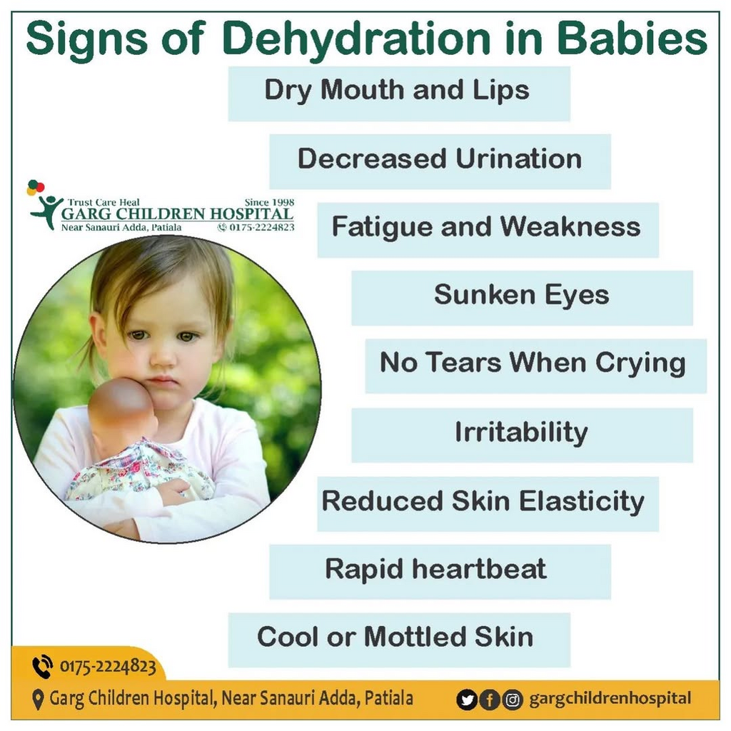
  • Hydration status:
    • Sunken eyes
    • Dry mucosa
    • Reduced urine output
  • Growth parameters (failure to thrive?)

Systemic Examination

Abdomen

  • Distension → obstruction
  • Tenderness → appendicitis
  • Mass → intussusception

CNS

  • Bulging fontanelle (infants)
  • Neck stiffness
  • Altered consciousness

Skin

  • Rash → infection/allergy
  • Petechiae → sepsis

Step 6: Investigations

👉 No “routine panel” exists — investigations should be targeted.

Basic Investigations

  • Serum electrolytes
  • Blood glucose
  • Blood gas (if severe)

When Indicated

  • Imaging
    • X-ray abdomen → obstruction
    • Ultrasound → intussusception
    • CT/MRI → CNS causes
  • Other tests
    • Urine analysis → UTI
    • LFT/RFT → systemic disease
    • Metabolic screening

Step 7: Management Approach

1. Treat the Cause (Definitive)

  • Surgery → obstruction
  • Antibiotics → infection
  • Insulin → DKA

2. Correct Dehydration (MOST IMPORTANT)

Mild–Moderate:

  • Oral Rehydration Therapy (ORT)

Severe:

  • IV fluids (bolus + maintenance)

3. Symptomatic Treatment

  • Ondansetron for persistent vomiting
  • NG decompression in obstruction
  • Electrolyte correction

4. Nutritional Support

  • Early feeding when tolerated
  • Continue breastfeeding

Step 8: Clinical Patterns to Recognize (Exam Gold)

PatternLikely Diagnosis
Projectile, non-biliousPyloric stenosis
Bilious vomitingIntestinal obstruction
Vomiting + diarrheaGastroenteritis
Vomiting + headache (morning)Raised ICP
Vomiting + abdominal pain → laterAppendicitis
Episodic vomiting, symptom-free intervalsCyclic vomiting

Common Pitfalls

  • Ignoring bilious vomiting
  • Missing appendicitis early
  • Assuming all vomiting = gastroenteritis
  • Not checking hydration status
  • Over-ordering unnecessary tests

Clinical Algorithm (Simple Mental Model)

  1. Is the child sick? (ABC + red flags)
  2. What is the age?
  3. What is the type of vomiting?
  4. What are associated symptoms?
  5. Targeted investigations
  6. Treat dehydration + cause

Key Takeaways

  • Most pediatric vomiting is benign and self-limiting
  • But always rule out life-threatening causes first
  • Age + vomiting type + red flags = diagnosis
  • Hydration management saves lives

Conclusion

Vomiting in children is a diagnostic puzzle—but a structured approach simplifies it. The goal is not to memorize hundreds of causes, but to quickly identify danger, localize the system involved, and act appropriately.

DEXTROSE INFUSION RATE (GIR) IN NEONATAL HYPOGLYCEMIA

1. Definition

GIR (Glucose Infusion Rate) = amount of glucose delivered per kg body weight per minute.

👉 Unit: mg/kg/min


2. Formula

GIR=Dextrose (%) ×Infusion rate (mL/kg/day)144GIR = \frac{\text{Dextrose (\%) } \times \text{Infusion rate (mL/kg/day)}}{144}GIR=144Dextrose (%) ×Infusion rate (mL/kg/day)​

👉 Alternate (commonly used in ICU):GIR=Dextrose (g/mL)×mL/kg/min×10001GIR = \frac{\text{Dextrose (g/mL)} \times \text{mL/kg/min} \times 1000}{1}GIR=1Dextrose (g/mL)×mL/kg/min×1000​


3. Normal Glucose Requirement (Term Neonate)

AgeGIR (mg/kg/min)
First 24 hrs4–6
After 24 hrs5–8

4. Treatment of Hypoglycemia

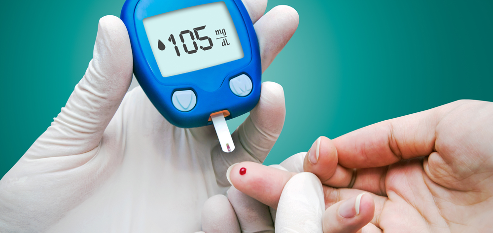
Symptomatic OR RBS <40 mg/dL

Step 1: Bolus

  • 10% Dextrose
  • Dose: 2 mL/kg IV bolus

Step 2: Continuous Infusion

SeverityStarting GIR
Mild4–6 mg/kg/min
Moderate6–8 mg/kg/min
Severe/refractory8–12 mg/kg/min

5. Escalation Strategy

  • Increase GIR by 2 mg/kg/min every 30–60 min
  • Max GIR: 12–15 mg/kg/min (sometimes up to 20 in refractory cases)

👉 If still hypoglycemic:

  • Consider:
    • Sepsis
    • Hyperinsulinism
    • Endocrine causes

6. Practical Conversion Table

GIR (mg/kg/min)D10 Rate (mL/kg/day)
460
575
690
8120
10150

👉 Quick memory:
D10 @ 80–100 mL/kg/day ≈ GIR 5–7


7. When High GIR Needed (>8–10)

Think:

  • Hyperinsulinism
  • Sepsis
  • Inborn errors of metabolism
  • Hypopituitarism
  • Adrenal insufficiency

8. Choice of Dextrose Concentration

FluidUse
D10First-line
D12.5Peripheral max
D15–D25Central line only

9. Monitoring

  • RBS every 30–60 min initially
  • Then every 4–6 hrs
  • Watch for:
    • Rebound hypoglycemia
    • Fluid overload

10. Weaning

  • Reduce GIR gradually once stable (>50–60 mg/dL)
  • Start/advance enteral feeds

Exam Pearls

  • First step = D10 bolus (2 mL/kg)
  • Target glucose >45 mg/dL
  • GIR >8 = suspect pathology
  • Max peripheral dextrose = 12.5%
  • Persistent hypoglycemia → think hyperinsulinism

Lumbar Puncture (LP) in Pediatrics & Neonates

1. Definition

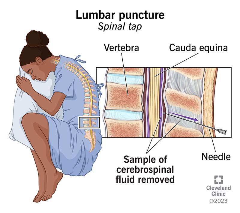
Lumbar puncture (LP) is a procedure in which a needle is inserted into the subarachnoid space of the lumbar spine to obtain cerebrospinal fluid (CSF) for diagnostic or therapeutic purposes.

Commonly done at L3–L4 or L4–L5 intervertebral space.


2. Indications

A. Diagnostic Indications

1. Suspected CNS infection

  • Meningitis
    • Bacterial
    • Viral
    • Tubercular
    • Fungal
  • Encephalitis
  • Brain abscess (supportive)
  • Neurosyphilis

2. Neonatal sepsis evaluation

Important in:

  • Neonates with positive blood culture
  • Sepsis with neurologic signs
  • Late onset sepsis
  • Persistent unexplained illness

3. Neurologic disorders

  • Guillain-Barré syndrome (albuminocytologic dissociation)
  • Multiple sclerosis
  • Demyelinating diseases
  • Leukodystrophy

4. Malignancy

  • Leukemia CNS involvement
  • Lymphoma
  • Medulloblastoma spread

5. Subarachnoid hemorrhage

When CT scan is negative but suspicion persists.


B. Therapeutic Indications

  • Intrathecal chemotherapy
  • Intrathecal antibiotics
  • Spinal anesthesia
  • CSF pressure reduction (rare)

3. Indications Specific to Neonates

Perform LP in neonates with:

  1. Suspected meningitis
  2. Positive blood culture
  3. Seizures
  4. Bulging fontanelle
  5. Apnea / unexplained respiratory deterioration
  6. Neurological abnormalities
  7. Late onset sepsis (>72 hrs)

May delay LP in unstable neonate until stabilization.


4. Contraindications

Absolute Contraindications

  • Signs of raised intracranial pressure with mass lesion
  • Cardiorespiratory instability
  • Local infection at puncture site

Relative Contraindications

  • Severe thrombocytopenia (<50,000)
  • Coagulopathy
  • Spinal deformity
  • Suspected spinal cord mass
  • Severe shock

5. Signs Suggesting Raised ICP (Do NOT perform LP immediately)

  • Focal neurological deficits
  • Papilledema
  • Altered consciousness
  • Unequal pupils
  • Abnormal posturing
  • Hypertension with bradycardia (Cushing triad)

These require neuroimaging first.


6. Anatomy

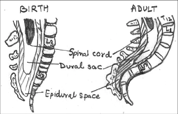
LP is done below the conus medullaris.

AgeConus level
NeonateL3
AdultL1–L2

Safe spaces:

  • L3–L4
  • L4–L5

Landmark:
Tuffier line (line joining iliac crests) → L4 vertebra


7. Equipment

  • Sterile gloves
  • Antiseptic solution
  • Sterile drapes
  • Spinal needle with stylet
  • Manometer
  • 3–4 sterile tubes
  • Local anesthetic (lidocaine)
  • Syringes
  • Adhesive dressing

8. Needle Size

AgeNeedle
Neonate22–25G
Infant22G
Child20–22G

Typical length:

  • Neonate: 1.5 inch
  • Older children: 2.5 inch

9. Position

1. Lateral decubitus (preferred)

  • Knees flexed to chest
  • Chin flexed
  • Allows opening pressure measurement

2. Sitting position

Used when landmarks difficult.


10. Procedure Steps

  1. Position child
  2. Identify L3–L4 or L4–L5
  3. Clean with antiseptic
  4. Sterile draping
  5. Local anesthesia
  6. Insert needle midline with stylet
  7. Advance slowly
  8. Feel “pop” entering subarachnoid space
  9. Remove stylet → CSF flows
  10. Collect CSF in tubes
  11. Replace stylet and remove needle

11. CSF Collection Tubes

TubeTest
Tube 1Biochemistry (protein, glucose)
Tube 2Microbiology (Gram stain, culture)
Tube 3Cell count
Tube 4Special tests (PCR, viral studies)

12. Opening Pressure

Measured with manometer.

Normal values:

AgePressure
Neonates2–6 cm H₂O
Children10–28 cm H₂O

13. Normal CSF Values

Neonates

ParameterNormal
Cells0–20/mm³
Protein40–120 mg/dL
Glucose2/3 blood glucose

Infants & Children

ParameterNormal
Cells0–5/mm³
Protein15–45 mg/dL
Glucose2/3 serum

14. CSF Interpretation

Bacterial Meningitis

FindingResult
Cells↑ (100–10,000)
Cell typeNeutrophils
Protein↑↑
Glucose↓
Opening pressure↑

Viral Meningitis

FindingResult
Cells10–1000
Cell typeLymphocytes
ProteinMild ↑
GlucoseNormal

TB Meningitis

FindingResult
Cells50–500
TypeLymphocytes
Protein↑↑
Glucose↓

15. Complications

Immediate

  • Traumatic tap
  • Pain
  • Bleeding
  • Infection

Post LP

  • Post-LP headache
  • Back pain

Serious

  • Brain herniation
  • Epidural hematoma
  • Nerve injury

16. Traumatic Tap Differentiation

FeatureTraumatic tapSAH
RBC countDecreases in later tubesSame in all tubes
ClotPresentAbsent
XanthochromiaAbsentPresent

17. Reasons for Failed LP

  • Wrong level
  • Poor positioning
  • Obesity
  • Dehydration
  • Needle obstruction

18. When NOT to Delay Antibiotics

In suspected meningitis:

  • Start antibiotics immediately
  • LP should not delay treatment

19. Special Considerations in Neonates

  • Higher CSF protein normally
  • CSF WBC slightly higher
  • LP often required in late onset sepsis
  • May be delayed if unstable

✅ Exam Pearls (Important for MD / Residency)

  • Best site: L3–L4
  • Neonatal normal CSF protein higher
  • Always replace stylet before removing needle
  • Opening pressure measured only in lateral position
  • Papilledema → neuroimaging before LP