Author Archives: CJ

About CJ

A clinician! Physician and a great doctor practicing for more than 5 years.

circle of willis

Circle of Willis: Discription and an Easy Mnemonic to Remember It

The Circle of Willis is a critical anatomical structure in the brain that plays a vital role in ensuring proper blood flow to this essential organ. Named after the English physician Thomas Willis, who first described it in the 17th century, this circular network of arteries is located at the base of the brain. It serves as a safety mechanism, providing collateral blood flow between the anterior and posterior circulations of the brain. Understanding its anatomy and function is crucial for medical professionals, students, and anyone interested in brain health. In this article, we’ll explore the Circle of Willis, its significance, and an easy mnemonic to help you remember its components.


What is the Circle of Willis?

The Circle of Willis is a ring-like structure formed by the interconnection of several major arteries. It ensures that blood supply to the brain remains consistent, even if one of the arteries becomes blocked or narrowed. This redundancy is vital for preventing strokes and other brain-related conditions.

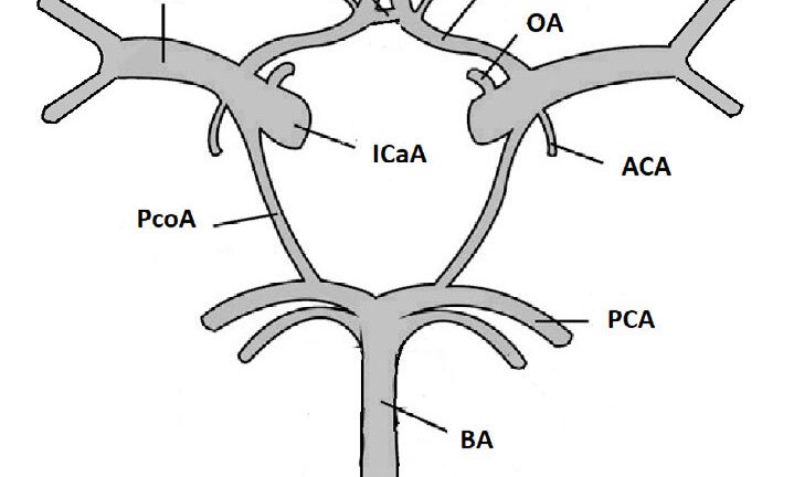
The primary arteries that make up the Circle of Willis include:

  1. Anterior Cerebral Arteries (ACA) – Supply blood to the frontal lobes of the brain.
  2. Anterior Communicating Artery (ACommA) – Connects the two anterior cerebral arteries.
  3. Internal Carotid Arteries (ICA) – Major arteries that supply blood to the brain.
  4. Posterior Cerebral Arteries (PCA) – Supply blood to the occipital lobes and parts of the temporal lobes.
  5. Posterior Communicating Arteries (PCommA) – Connect the internal carotid arteries to the posterior cerebral arteries.
  6. Basilar Artery – Formed by the union of the two vertebral arteries, it supplies blood to the brainstem and cerebellum.

Why is the Circle of Willis Important?

The Circle of Willis acts as a backup system for blood flow. If one artery becomes blocked due to a clot or narrowing, blood can still reach the affected area through alternative pathways within the circle. This collateral circulation is essential for maintaining brain function and preventing ischemic strokes.

However, not everyone has a complete Circle of Willis. Variations in its anatomy are common, and some people may have missing or underdeveloped arteries. These variations can affect how well the brain compensates for reduced blood flow.


An Easy Mnemonic to Remember the Circle of Willis

Memorizing the components of the Circle of Willis can be challenging, but mnemonics can make it easier. Here’s a simple and effective mnemonic to help you remember the key arteries:

“A Cat In The House Can Purr Always”

Let’s break it down:

  • A – Anterior Cerebral Arteries (ACA)
  • Cat – Anterior Communicating Artery (ACommA)
  • In – Internal Carotid Arteries (ICA)
  • The – Posterior Communicating Arteries (PCommA)
  • House – Posterior Cerebral Arteries (PCA)
  • Can – (Basilar Artery is implied as the central connection)
  • Purr Always – (Helps reinforce the mnemonic)

This mnemonic simplifies the complex anatomy of the Circle of Willis, making it easier to recall during exams or clinical practice.


Clinical Significance of the Circle of Willis

The Circle of Willis is not just an anatomical curiosity; it has significant clinical implications. For example:

  1. Stroke Prevention: A well-developed Circle of Willis can reduce the risk of stroke by providing alternative blood flow pathways.
  2. Aneurysms: Weak spots in the arteries of the Circle of Willis can lead to aneurysms, which are life-threatening if they rupture.
  3. Surgical Planning: Understanding the anatomy of the Circle of Willis is crucial for neurosurgeons when planning procedures like bypass surgeries or treating aneurysms.

Conclusion

The Circle of Willis is a fascinating and vital structure in the human brain, ensuring a steady supply of blood to this critical organ. Its role in maintaining brain health cannot be overstated, and understanding its anatomy is essential for medical professionals and students alike. With the help of the mnemonic “A Cat In The House Can Purr Always,” remembering the components of the Circle of Willis becomes much simpler.

Whether you’re studying for an exam, preparing for a medical career, or simply curious about how your brain works, the Circle of Willis is a topic worth exploring. By appreciating its importance, we can better understand the complexities of brain health and the incredible design of the human body.

Screening vs diagnostic tests : 12 major differences and top important MCQs

Introduction

Screening tests and diagnostic tests serve different purposes in healthcare:

  • Screening tests are used to identify potential disease in asymptomatic individuals. They are usually less invasive, widely applied, and meant to detect early signs of disease. A positive result often requires further diagnostic testing.
    • Example: Mammography for breast cancer screening.
  • Diagnostic tests are used to confirm the presence of a disease in individuals who have symptoms or a positive screening result. They are typically more specific and may be more invasive or expensive.
    • Example: Biopsy to confirm breast cancer after an abnormal mammogram.

In short, screening detects risk, while diagnosis confirms disease.

Table of differences Screening vs Diagnostic tests

Feature Screening Test Diagnostic Test
Purpose Detects potential disease in asymptomatic individuals Confirms disease in symptomatic individuals or those with positive screening results
Target Population Large, general population at risk Individuals with symptoms or positive screening results
Timing Performed before symptoms appear Performed after symptoms appear or after a positive screening test
Outcome Indicates the need for further testing Provides a definitive diagnosis
Invasiveness Usually non-invasive Can be invasive
Cost Generally lower Often more expensive
Accuracy High sensitivity (to detect most cases) but may have false positives High specificity (to confirm disease with accuracy)
False Positives/Negatives More false positives (may identify healthy people as at risk) Lower false positives but higher false negatives in some cases
Follow-up Needed? Yes, a positive result requires further diagnostic testing No, provides a final diagnosis
Repetition Performed at regular intervals (e.g., yearly) Done once or as needed for confirmation
Population-Based or Individual-Based? Population-based Individual-based
Example – Cancer Mammogram for breast cancer screening Biopsy to confirm breast cancer
Example – Infectious Disease HIV rapid test Western blot or PCR test for HIV confirmation
Example – Pregnancy Home pregnancy test (detects hCG) Ultrasound to confirm pregnancy location and viability

MCQs on Screening vs. Diagnostic Tests

1. What is the primary purpose of a screening test?

A) To confirm the presence of a disease
B) To detect potential disease in asymptomatic individuals
C) To provide treatment for a disease
D) To identify the severity of a disease

2. Which of the following is an example of a screening test?

A) Biopsy for cancer
B) Blood glucose test for diabetes symptoms
C) Mammogram for breast cancer in healthy individuals
D) MRI scan for severe headaches

3. Screening tests are usually performed on which type of population?

A) Only symptomatic individuals
B) Only individuals with family history of a disease
C) General population at risk but without symptoms
D) Patients already diagnosed with a disease

4. How do screening tests differ from diagnostic tests in terms of cost and invasiveness?

A) Screening tests are more expensive and invasive
B) Diagnostic tests are less expensive and non-invasive
C) Screening tests are usually less expensive and non-invasive
D) There is no difference in cost and invasiveness between the two

5. What happens if a screening test result is positive?

A) The patient is immediately treated for the disease
B) The test confirms the disease without further evaluation
C) Further diagnostic testing is required to confirm the disease
D) No further action is needed

6. Which of the following characteristics is typical of a diagnostic test?

A) Performed on asymptomatic individuals
B) Typically repeated at regular intervals
C) Highly specific and confirms disease presence
D) Conducted on a large population without symptoms

7. Which of the following is NOT a feature of screening tests?

A) High sensitivity to detect most cases
B) Regularly performed at set intervals
C) Used to confirm a definitive diagnosis
D) Conducted on a wide population

8. Which of the following screening tests is used for infectious diseases?

A) HIV rapid test
B) Biopsy
C) MRI
D) Ultrasound

9. Which statement about false positives in screening tests is true?

A) Screening tests rarely produce false positives
B) False positives can lead to unnecessary follow-up tests
C) False positives are not possible in any medical test
D) False positives confirm the presence of a disease

10. A diagnostic test is typically performed:

A) On all individuals, regardless of symptoms
B) On people showing symptoms or with a positive screening test
C) Only when no symptoms are present
D) To replace screening tests


Answer Key

  1. B – To detect potential disease in asymptomatic individuals
  2. C – Mammogram for breast cancer in healthy individuals
  3. C – General population at risk but without symptoms
  4. C – Screening tests are usually less expensive and non-invasive
  5. C – Further diagnostic testing is required to confirm the disease
  6. C – Highly specific and confirms disease presence
  7. C – Used to confirm a definitive diagnosis
  8. A – HIV rapid test
  9. B – False positives can lead to unnecessary follow-up tests
  10. B – On people showing symptoms or with a positive screening test

10 model questions for the Nepal Nursing Council (NNC) Licensing Exam 2081

1. Which of the following is the most appropriate nursing intervention for a patient experiencing hypoglycemia?

A) Administer intravenous insulin
B) Give a glass of fruit juice or glucose orally
C) Encourage the patient to exercise
D) Monitor blood glucose after 24 hours

Key: B) Give a glass of fruit juice or glucose orally


2. A patient with chronic obstructive pulmonary disease (COPD) is on oxygen therapy. What is the recommended oxygen flow rate to prevent oxygen toxicity?

A) 1-2 L/min
B) 4-6 L/min
C) 6-8 L/min
D) 10-12 L/min

Key: A) 1-2 L/min


3. Which of the following signs and symptoms are most commonly associated with right-sided heart failure?

A) Pulmonary congestion and dyspnea
B) Peripheral edema and jugular vein distension
C) Hypertension and headache
D) Chest pain and palpitations

Key: B) Peripheral edema and jugular vein distension


4. A nurse is assessing a patient with meningitis. Which of the following clinical signs is indicative of meningeal irritation?

A) Kernig’s sign
B) Trousseau’s sign
C) Chvostek’s sign
D) Babinski’s sign

Key: A) Kernig’s sign


5. The primary goal of nursing care for a patient in the immediate postoperative period is to:

A) Ensure the patient’s pain is completely eliminated
B) Monitor for complications and maintain airway patency
C) Encourage early ambulation to prevent bedsores
D) Administer IV fluids to prevent dehydration

Key: B) Monitor for complications and maintain airway patency


6. A patient receiving digoxin therapy is experiencing nausea, vomiting, and visual disturbances. What should the nurse do first?

A) Administer an antiemetic
B) Check the patient’s serum potassium level
C) Hold the medication and notify the physician
D) Encourage the patient to drink more water

Key: C) Hold the medication and notify the physician


7. Which of the following precautions should be followed while administering intramuscular injections in children under 2 years of age?

A) Use the dorsogluteal site
B) Use the deltoid muscle
C) Use the vastus lateralis muscle
D) Use a 3-inch needle

Key: C) Use the vastus lateralis muscle


8. A patient with tuberculosis is started on rifampicin therapy. Which of the following instructions should the nurse give?

A) Stop the medication if urine turns orange
B) Take the medication with milk
C) Avoid alcohol while on this medication
D) This medication is only needed for two weeks

Key: C) Avoid alcohol while on this medication


9. Which of the following is an early sign of increased intracranial pressure (ICP)?

A) Bradycardia
B) Hypertension
C) Altered level of consciousness
D) Fixed and dilated pupils

Key: C) Altered level of consciousness


10. A nurse is caring for a patient with a deep vein thrombosis (DVT) in the left leg. Which intervention should be avoided?

A) Elevating the affected leg
B) Applying warm compresses
C) Encouraging bed rest
D) Massaging the affected leg

Key: D) Massaging the affected leg


Explanation of NNC license model Questions

  1. Hypoglycemia Management
    Rationale: Hypoglycemia (low blood sugar) is treated with quick-acting carbohydrates, such as fruit juice or glucose tablets, to rapidly raise blood sugar levels. Insulin administration (option A) would worsen the condition.
  2. Oxygen Therapy in COPD
    Rationale: In COPD patients, high oxygen levels can suppress the respiratory drive. A low-flow oxygen rate (1-2 L/min) is recommended to prevent carbon dioxide retention and respiratory failure.
  3. Right-Sided Heart Failure
    Rationale: Right-sided heart failure leads to fluid retention and systemic venous congestion, causing peripheral edema (swelling) and jugular vein distension (JVD) due to increased pressure in the venous system.
  4. Meningitis Signs
    Rationale: Kernig’s sign (pain and resistance with leg extension when the hip is flexed) is a classic indication of meningeal irritation seen in bacterial or viral meningitis.
  5. Postoperative Nursing Priorities
    Rationale: The most critical concern after surgery is maintaining airway patency and preventing complications such as respiratory distress, bleeding, or shock. Pain control is important but secondary to airway management.
  6. Digoxin Toxicity
    Rationale: Digoxin toxicity presents with nausea, vomiting, visual disturbances (yellow vision), and bradycardia. The nurse should hold the drug and notify the physician to prevent severe toxicity and cardiac complications.
  7. IM Injection in Children
    Rationale: The vastus lateralis muscle (thigh) is the safest site for intramuscular injections in infants and young children because it has a large muscle mass and fewer major nerves or blood vessels.
  8. Rifampicin Therapy in Tuberculosis
    Rationale: Rifampicin (used in TB treatment) can cause orange-colored urine (a harmless side effect), but alcohol should be avoided as it increases liver toxicity.
  9. Increased Intracranial Pressure (ICP)
    Rationale: The earliest sign of increased ICP is an altered level of consciousness, including confusion or drowsiness, due to brain pressure affecting neurological function. Late signs include bradycardia and dilated pupils.
  10. Deep Vein Thrombosis (DVT) Precautions
    Rationale: Massaging a DVT-affected leg can dislodge a clot, potentially leading to a pulmonary embolism, which can be life-threatening. Instead, elevating the leg and ensuring proper blood circulation is recommended.

How To Measure Waist Hip Circumference Ratio: 5 easy ways to control waist hip circumference ratio

Definition of Waist-to-Hip Ratio (WHR) (waist hip circumference ratio)

Waist-to-Hip Ratio (WHR) is the ratio of the circumference of the waist to that of the hips. It is calculated by dividing the waist measurement by the hip measurement. WHR is an indicator of body fat distribution and is used as a measure of health risk associated with obesity.

WHR (waist hip circumference ratio) Classification

The WHR classification is as follows:

risk level Male female
lower risk less than 0.9 less than 0.8
moderate risk 0.92-0.99 0.82-0.84
high risk more than equal to 1 more than equal to 0.85

A higher WHR indicates greater central fat distribution, which is associated with an increased risk of cardiovascular disease, type 2 diabetes, and other obesity-related conditions.

Technique of measuring waist and hip circumference

Waist Circumference

Measure the smallest circumference of the abdomen, just above the belly button (navel), after exhaling naturally.

Waist hip citcumference ratio

Hip Circumference

Measure the largest circumference of the hips, typically around the widest part of the buttocks.

The waist and hip circumference ratio calculator

Calculate Your Waist-to-Hip Ratio (WHR)


Result:

How to Reduce Waist and Hip Circumference

1. Balanced Diet: Eat a diet rich in fruits, vegetables, lean protein, and whole grains while avoiding excessive sugar and processed foods.

2. Regular Exercise:

  • Engage in aerobic exercises (e.g., running, swimming, cycling) to burn fat.
  • Incorporate strength training to build muscle and improve metabolism.

3. Hydration: Drink plenty of water to support digestion and metabolism.

4. Adequate Sleep: Aim for 7-9 hours of sleep per night to regulate hormones that control appetite and fat storage.

5. Stress Management: Practice mindfulness, meditation, or yoga to manage stress and avoid emotional eating.

FAQs

1. What is Waist-to-Hip Ratio (WHR), and why is it important?

WHR is the size of your waist divided by the size of your hips. It tells how fat is stored in your body. A high WHR can mean a higher risk of heart problems, diabetes, and other health issues.

2. How do I measure my waist and hips correctly?

For your waist, measure just above your belly button (the narrowest part of your abdomen).
For your hips, measure around the widest part of your hips and buttocks.
Use a tape measure, and make sure it’s snug but not too tight.

3. How can I lower my waist-to-hip ratio?

Eat healthy foods like fruits, vegetables, and lean proteins.
Exercise regularly (cardio and strength training).
Drink enough water, sleep well, and reduce stress.
These steps can help reduce fat, especially around your waist, and improve your overall health.

Top 5 Facts about Gower’s Sign: How to easily demonstrate Gower’s sign?

Gowers’ sign is a medical sign that indicates weakness in the pelvic girdle and proximal lower limb muscles. It’s characterized by a patient using their hands to “walk” up their body to get to a standing position. 

10 red flags in a relationship that you should never ignore?

How is Gower’s Sign Performed?

  1. Start in a supine or sitting position
  2. Roll onto your stomach with your arms and legs extended
  3. Put your hands on the ground and shift your weight onto your extended arms
  4. Push your body backward to extend your legs
  5. Put your hands on your knees
  6. Walk your hands up your thighs until you’re standing

What it can indicate

  • Duchenne muscular dystrophy: A characteristic sign of this condition 
  • Guillain-Barré syndrome (GBS): Can be a sign of recovery from GBS 
  • Pelvic girdle weakness: A sign of weakness in the pelvic girdle and proximal lower extremity muscles 
  • Diskitis: Can be associated with diskitis at the L4-5 intervertebral space 

Who described it?

Gowers’ sign was described by neurologist Sir William Richard Gowers in 1879. 

Now lets dig into DMD.

Read now: Why is the MDF Stethoscope today’s No 1 Choice as Medical Professionals?

Duchenne Muscular Dystrophy (DMD)

Duchenne Muscular Dystrophy (DMD) is the most common and severe form of progressive muscular dystrophy in children. It belongs to a group of primary myopathies characterized by a progressive, genetic-based degeneration and death of muscle fibers.

1. Genetic Profile and Pathogenesis

  • Inheritance: DMD is an X-linked recessive disorder, almost exclusively affecting males and carried by females. Approximately one-third of cases result from de novo (new) mutations.
  • Molecular Defect: It is caused by a mutation (typically a deletion) in the gene for dystrophin located on chromosome Xp21.
  • Mechanism: Dystrophin is the longest known human gene and encodes a protein that connects the muscle fiber cytoskeleton to the surrounding extracellular matrix. Without functional dystrophin, muscle cells undergo chronic damage, necrosis, and replacement by fat and fibrous tissue.

2. Clinical Features

DMD typically presents between the ages of 2 and 5 years.

  • Early Signs: Delayed motor milestones (especially late walking) and mild speech or language delay are often the first indicators.
  • Muscle Weakness: Progressive, symmetric weakness begins in the pelvis and hip girdle, later involving the shoulder girdle.
  • Gait and Mobility: Affected children exhibit a waddling (Trendelenburg) gait, frequent falls, and difficulty climbing stairs (mounting them “one by one”).
  • Gower Sign: A classic clinical finding where the child, unable to stand up normally from the floor, must turn prone and “climb up his own thighs” with his hands to reach an upright position.
  • Pseudohypertrophy: Striking enlargement of the calf muscles occurs because muscle tissue is replaced by fat and connective tissue rather than true muscle growth.
  • Intellectual Function: Approximately 20% to 30% of boys have learning difficulties or non-progressive intellectual impairment.

3. Diagnostic Evaluation

Diagnosis should be initiated at the first clinical suspicion.

  • Serum Creatine Kinase (CK): Remarkably high levels (usually 10 to 40 times normal) are a hallmark of the disease.
  • Genetic Testing: Direct dystrophin gene testing is now the preferred definitive method and often avoids the need for a muscle biopsy.
  • Electromyography (EMG): Shows a “myopathic” pattern, including reduced amplitude and duration of motor unit potentials.
  • Muscle Biopsy: If performed, shows fiber necrosis, phagocytosis, and increased endomysial fat and connective tissue.

4. Progression and Complications

  • Loss of Ambulation: Most boys become wheelchair-bound by age 10 to 13.
  • Orthopedic: As weakness progresses, many develop scoliosis, particularly once they are no longer walking.
  • Cardiac: Progressive cardiomyopathy and persistent tachycardia develop in nearly all patients.
  • Respiratory: Weakness of the diaphragm and intercostal muscles leads to nocturnal hypoxia and eventual respiratory failure.
  • Prognosis: Death typically occurs in the late teens or 20s from cardiorespiratory complications.

5. Management

Management requires a specialist multidisciplinary team.

  • Corticosteroids: (e.g., Prednisone or Deflazacort) are the mainstay of treatment, as they preserve mobility, improve muscle strength, and prevent scoliosis.
  • Supportive Care: Physiotherapy and splinting are used to prevent contractures. Nocturnal hypoxia may be managed with overnight CPAP.
  • Novel Therapies: Research into exon-skipping drugs (e.g., Ataluren or Eteplirsen) allows for the production of small amounts of functional dystrophin in patients with specific mutations.
  • Genetic Counseling: Crucial for the family to detect female carriers and discuss antenatal diagnosis.

DMD vs. Becker Muscular Dystrophy (BMD): BMD is a milder allelic variant where some functional dystrophin is produced. BMD features a later onset (average 11 years), a slower course, and patients typically remain ambulatory into their late 20s or beyond, with many living into middle age.

Who described Gower’s sign?

Sir William Richard Gowers

What can Gower’s Sign indicate?

Duchenne muscular dystrophy, Guillain-Barré syndrome (GBS), pelvic girdle weakness, and diskitis. 

18 Top Funny African Proverbs You Must Read Now

Proverb related to beehives

When a man is stung by a bee, he does not destroy all beehives- Kenya Proverb

Proverb related to stupid

It is only a stupid cow that rejoices at the prospect of being taken to a beautiful abattoir

Zambian Proverb

Proverb related to eagle

No matter how far an eagle flies up the sky, it will definitely come down to look for food

– Zimbabwe Proverb

Proverb related to sheep

An army of sheep led by a lion can defeat an army of lions led by a sheep.

Ghanaian proverb

Proverb related to anus

The anus doesn’t teach the mouth the sweetness of food

– South-African Proverb

Proverb related to woman

The man who marries a beautiful woman and the farmer who grows corn by the roadside have the same problem

Ethiopian Proverb

Proverb related to man

A short man is not a boy.

– Nigerian Proverb

Proverb related to anger

No matter how hot your anger is, it cannot cook yam.

– Nigerian Proverb

Proverb related to carefulness

It requires a lot of carefulness to kill the fly that perches on the scrotum

Ghanaian Proverb

Proverb related to throat

If the throat can grant passage to a knife, the anus should wonder how to expel it

-South-African Proverb

Proverb related to frown on the face

The frown on the face of the goat will not stop it from being taken to the market

– Nigerian Proverb

Proverb related to old lady

An old lady feels uneasy when dry bones are mentioned in a proverb

Ghanaian Proverb

Proverb related to wax

The same sun that melts the wax hardens the clay

Nigerien Proverb

Proverb related to road

If you don’t know where you are going, any road will take you there

– Ugandan Proverb

Proverb related to virgin

There is no virgin in a maternity ward

– Cameroonian Proverb

Proverb related to madman

The madman, who throws a stone into a crowded market, forgets that his own mother could be hit by his madness.

– Ugandan Proverb

Proverb related to mother’s breasts

A child can play with its mother’s breasts, but not its father’s testicles. – Guinean Proverb

Proverb related to itching anus

He who goes to sleep with an itching anus wakes up with smelly fingers – Nigerian Proverb