Category Archives: Health

skeletal muscle

Creatine Kinase (CK) – High-Yield Medical Notes

1. Definition

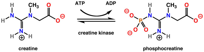
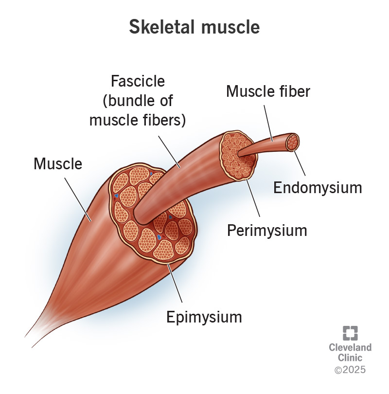
Creatine kinase (CK), also called creatine phosphokinase (CPK), is an intracellular enzyme that catalyzes the reversible conversion:Creatine+ATP↔Phosphocreatine+ADPCreatine + ATP \leftrightarrow Phosphocreatine + ADPCreatine+ATP↔Phosphocreatine+ADP

This reaction is crucial for energy storage and rapid regeneration of ATP, especially in muscle and brain tissues.


2. Location in Body

CK is present mainly in tissues with high energy demand:

TissueCK concentration
Skeletal muscleHighest
Cardiac muscleHigh
BrainModerate
Smooth muscleLow

Because CK is intracellular, serum CK rises when cell membrane damage occurs.


3. CK Isoenzymes

CK exists in three isoenzymes, formed by combinations of M (muscle) and B (brain) subunits.

IsoenzymeStructureMajor SourceClinical significance
CK-MMM + MSkeletal muscleMuscle injury, rhabdomyolysis
CK-MBM + BCardiac muscleMyocardial injury
CK-BBB + BBrain, smooth muscleCNS injury

Distribution

  • CK-MM: ~95–100% of total CK in normal serum
  • CK-MB: <5% normally
  • CK-BB: normally absent in serum

4. Normal Values

Normal ranges vary by lab.

Typical reference:

GroupCK (U/L)
Adult male50–200
Adult female40–150
ChildrenHigher than adults
NeonatesCan be very high after birth

5. Causes of Elevated CK

A. Skeletal Muscle Disorders

Most common cause.

Examples:

  • Muscular dystrophy (e.g., Duchenne muscular dystrophy)
  • Inflammatory myopathies
    • Polymyositis
    • Dermatomyositis
  • Trauma
  • Intramuscular injections
  • Vigorous exercise

B. Rhabdomyolysis

Massive CK elevation.

Causes:

  • Crush injury
  • Drugs (statins)
  • Heat stroke
  • Severe infections

CK may rise >5000–10,000 U/L.


C. Cardiac Causes

CK-MB rises in myocardial injury.

Example:

  • Myocardial Infarction

However, CK-MB is now largely replaced by troponins.


D. Neurologic Disorders

CK-BB may increase in:

  • Stroke
  • Brain trauma
  • Seizures

Example:

  • Stroke

E. Other Causes

  • Hypothyroidism
  • Alcohol abuse
  • Surgery
  • Prolonged immobilization
  • Severe infections

6. CK in Myocardial Infarction

Historically important marker.

ParameterCK-MB
Rise3–6 hours
Peak12–24 hours
Normalization48–72 hours

Used previously to detect reinfarction.

Now replaced mainly by troponin I/T.


7. CK in Pediatric Practice

Important uses:

Screening for muscular dystrophy

Very high CK in:

  • Duchenne muscular dystrophy
  • Becker muscular dystrophy

Levels may be 10–100× normal.


Evaluation of Hypotonia

Used when evaluating:

  • floppy infant
  • muscle weakness

Evaluation of Rhabdomyolysis

Symptoms:

  • myalgia
  • weakness
  • dark urine (myoglobinuria)

8. Causes of Decreased CK

Rare clinical significance.

Seen in:

  • Low muscle mass
  • Chronic illness
  • Pregnancy

9. Drugs Causing Elevated CK

Important clinically.

Examples:

DrugMechanism
StatinsMyopathy
AntipsychoticsNeuroleptic malignant syndrome
CorticosteroidsMuscle breakdown

Example condition:
Neuroleptic Malignant Syndrome


10. Investigation Panel When CK is Elevated

TestPurpose
CK-MBCardiac source
TroponinMI confirmation
AST / ALTMuscle vs liver
LDHTissue injury
Serum myoglobinRhabdomyolysis
Urine myoglobinKidney injury

11. CK in Rhabdomyolysis Severity

CK levelInterpretation
<1000Mild
1000–5000Moderate

5000 | Severe muscle injury |
15000 | High risk of renal failure |


12. Clinical Pearls (Exam High-Yield)

  • CK-MM → skeletal muscle injury
  • CK-MB → cardiac muscle
  • CK-BB → brain
  • Duchenne muscular dystrophy → CK extremely high
  • CK rises after muscle trauma/exercise
  • CK-MB replaced by troponin in MI diagnosis

One-line exam memory tip

“CK rises with muscle damage — skeletal (MM), cardiac (MB), brain (BB).”

Aphthous Ulcers: Step-by-Step Workup and Management

1. Clinical Assessment

History

  • Onset, duration, frequency, and number of ulcers
  • Pain severity, triggers (trauma, stress, certain foods)
  • Systemic symptoms: fever, diarrhea, joint pain, fatigue
  • Family history of similar lesions
  • Medications (NSAIDs, beta-blockers)
  • Previous treatments and response

Examination

  • Location: non-keratinized mucosa (buccal, labial, ventral tongue, floor of mouth)
  • Size:
    • Minor (<1 cm) – most common
    • Major (>1 cm, deeper, may scar)
    • Herpetiform (multiple small ulcers)
  • Number: single or multiple
  • Appearance: round/oval, yellow-white pseudomembrane with erythematous halo
  • Rule out: secondary causes (herpes simplex, trauma, systemic disease)

2. Investigations (If atypical or recurrent)

  • Basic labs: CBC, iron studies, vitamin B12, folate (nutritional deficiencies)
  • CRP/ESR: if systemic symptoms
  • Serology: celiac disease (tTG-IgA), HIV (if risk factors)
  • Biopsy: rare, only if persistent >3 weeks, atypical, or suspected malignancy
  • Microbiology: rarely needed; ulcers are typically non-infectious

3. Classification

TypeFeaturesManagement Focus
Minor<1 cm, 1–5 lesions, heal 7–14 daysSymptomatic relief
Major>1 cm, deep, persistent, may scarSystemic therapy possible
HerpetiformMultiple, small, coalescing, painfulCombination topical/systemic therapy

4. Management

A. General Measures

  • Avoid triggers: spicy/acidic foods, trauma
  • Good oral hygiene
  • Avoid sodium lauryl sulfate in toothpaste
  • Stress management
  • Nutritional supplementation if deficient

B. Topical Therapy (First-line for minor ulcers)

  • Topical corticosteroids:
    • Triamcinolone acetonide 0.1% in orabase
    • Fluocinonide 0.05% gel
  • Topical anesthetics:
    • Lidocaine 2% gel for pain relief
  • Protective agents:
    • Orabase, hydroxypropyl cellulose films

C. Systemic Therapy (For severe, recurrent, or major ulcers)

  • Oral corticosteroids:
    • Prednisone short course (e.g., 30–60 mg/day tapering)
  • Immunomodulators (refractory cases):
    • Colchicine, thalidomide, dapsone
  • Address underlying systemic disease:
    • Nutritional deficiencies, IBD, Behçet’s disease, etc.

D. Adjunctive Therapy

  • Antimicrobial mouth rinses: chlorhexidine gluconate
  • Pain control: NSAIDs (if not contraindicated)
  • Laser therapy (experimental for pain reduction and healing)

5. Follow-Up

  • Minor ulcers: usually self-limiting; review if >3 weeks
  • Recurrent or major ulcers: monitor frequency, severity, and response to therapy
  • Evaluate for systemic disease if persistent or atypical

APGAR Score – An easy Quick Guide for Newborn Assessment


Introduction

The APGAR score is a rapid method to assess the health status of a newborn immediately after birth.
It was introduced in 1952 by Dr. Virginia Apgar, an American anesthesiologist, to provide an objective and standardized evaluation.

The score is based on five parameters, each evaluated at 1 minute and 5 minutes after birth, with each parameter scored from 0 to 2. The maximum score is 10.


APGAR Parameters

LetterParameter0 Points1 Point2 Points
AAppearance (Skin Color)Entire body bluish/cyanoticPink body, bluish hands & feet (acrocyanosis)Entire body pink
PPulse (Heart Rate)Absent< 100 bpm≥ 100 bpm
GGrimace (Reflex Response)No response to stimulationGrimace onlyCough, sneeze, or cry
AActivity (Muscle Tone)Limbs extended (flaccid)Some flexion of limbsActive motion, full flexion
RRespiratory EffortAbsentWeak cry or labored breathingStrong cry, good breathing

Interpreting the APGAR Score

  • 7–10: Normal, healthy newborn
  • 4–6: Mild depression (needs observation and possibly mild intervention)
  • 0–3: Severe depression (needs urgent medical attention)

When to Measure

  • 1-minute APGAR: Reflects the newborn’s condition at birth and initial adaptation to the environment.
  • 5-minute APGAR: Evaluates the effectiveness of ongoing care and resuscitation efforts.
  • If the 5-minute score is < 7, assessment should be repeated every 5 minutes up to 20 minutes.

Practical Note for Delivery Rooms

  • Look & Listen: Appearance, activity, respiratory effort
  • Feel: Heart rate, reflex response (grimace)

This quick check can be done within seconds and helps guide immediate care for the newborn.


Summary:
The APGAR score remains one of the simplest and most effective tools for assessing newborn well-being. It does not predict long-term outcomes but is essential for guiding immediate care and determining if urgent intervention is required.


What is freeze indicator in vaccine? A guide


1. Definition

A Freeze Indicator is a device or label used to detect whether a vaccine (or other temperature-sensitive product) has been exposed to temperatures at or below its freezing point—usually 0°C or lower.

  • Purpose: To prevent use of vaccines that have lost potency or safety due to freezing.
  • Target vaccines: Mainly freeze-sensitive vaccines containing aluminum adjuvant (e.g., DTP, Pentavalent, Hep B, HPV), which lose potency irreversibly when frozen.

2. Types of Freeze Indicators

TypeFunctionExample Devices
Single-use chemical indicatorsChange color irreversibly after exposure to freezing temperature for a certain durationFreeze-tag®
Electronic data loggersRecord continuous temperature; can be set to trigger an alarm if freezing occursLogTag®, ELPRO
Phase-change indicatorsContain material that solidifies/melts at 0°C, showing visible changeSome cold chain box indicators

3. Working Principle

  • Based on phase change of a material or thermochemical reaction triggered by freezing temperatures.
  • The change is irreversible, serving as a permanent record of freezing exposure.

4. Interpretation (Using Freeze-tag® example)

  • OK: Indicator window shows ✓ (no freezing exposure).
  • ALARM / ❌: Indicator window shows a cross mark (freezing detected).
  • Device cannot be reset—once tripped, the product must be considered potentially damaged.

5. Pediatric Relevance – Vaccines Affected by Freezing

  • DTP / DT / Td
  • Pentavalent
  • Hepatitis B
  • HPV
  • Pneumococcal conjugate vaccine (PCV)
  • Inactivated polio vaccine (IPV)
  • Hib (liquid formulations)

Freeze-stable vaccines (e.g., OPV, measles, BCG before reconstitution) are not affected by freezing.


6. Field Handling Notes

  • Always check freeze indicator + VVM before using a vaccine.
  • Avoid placing freeze-sensitive vaccines in direct contact with ice packs.
  • In outreach, use conditioned ice packs (sweating surface, internal ice still solid).
  • If freeze indicator shows alarm → vaccine should be discarded as per national guidelines.

7. Limitations

  • Some indicators respond only to certain freezing durations (e.g., >60 min below 0°C).
  • Cannot detect multiple freeze–thaw cycles unless electronic logger is used.
  • Doesn’t measure heat exposure—must be paired with VVM for complete cold chain safety.

If you want, I can make you a side-by-side pediatric cold chain safety chart comparing VVMs vs Freeze Indicators with examples, WHO limits, and affected vaccines so it’s all in one quick-reference sheet.
Would you like me to prepare that?

Vaccine vial Monitoring: Note for Pediatrician


1. Definition

A Vaccine Vial Monitor (VVM) is a small thermochromic label placed on a vaccine vial, ampoule, or dropper to monitor cumulative heat exposure over time.

  • Purpose: Helps ensure that vaccines have not been damaged by excessive heat during storage or transport in the cold chain.
  • Function: Color changes irreversibly with time and temperature.

2. Components & Principle

  • Center Square: Heat-sensitive material that darkens progressively with heat exposure.
  • Outer Reference Ring: Fixed color for comparison.
  • Working Principle:
    • Heat causes a chemical reaction in the center square.
    • The higher the temperature, the faster the change.
    • Based on Arrhenius kinetics—reaction rate doubles with ~10 °C rise in temperature.

3. Reading a VVM

  • Acceptable: If center square is lighter than outer ring → vaccine usable.
  • Discard: If center square is same or darker than outer ring → vaccine compromised.

4. VVM Types & Vaccine Shelf Life

WHO assigns VVM categories depending on vaccine heat stability:

VVM TypeTime to end-point at 37 °CExamples
VVM30≥ 30 daysOral polio (OPV), Hep B
VVM14≥ 14 daysDTP, Pentavalent
VVM7≥ 7 daysRotavirus
VVM2≥ 2 daysCertain lyophilized vaccines (e.g., measles, BCG before reconstitution)

5. Storage & Handling Notes for Pediatric Use

  • Check VVM before every use—especially in outreach/immunization camps.
  • Do not refrigerate below recommended temperature just to “reset” VVM—it’s irreversible.
  • VVM is not a freeze indicator—separate freeze indicators are used for freeze-sensitive vaccines (e.g., DTP, Hep B).
  • Post-reconstitution: VVM is invalid for multi-dose lyophilized vaccines—time limit per WHO multi-dose policy applies.

6. Field Significance in Pediatrics

  • Ensures safe vaccines for children in peripheral settings where cold chain breaches are common.
  • Reduces wastage by allowing use of vaccines that have been out of refrigeration but still within VVM limit.
  • Prevents administration of heat-damaged vaccines, which could cause reduced immunogenicity without visible signs.

7. Limitations

  • Cannot detect freezing damage.
  • Not a substitute for proper cold chain monitoring (data loggers, ice packs, cold boxes).
  • Only indicates cumulative heat exposure—not precise real-time temperature.

Why Do Female Astronauts Take Birth Control Pills in Space? The Surprising Science Behind It

Space travel is a marvel of human ingenuity, a daring leap into the unknown. But beyond the sleek spacecraft and zero-gravity acrobatics, astronauts face a very human challenge: their own biology. For female astronauts, one of the lesser-known but fascinating aspects of spaceflight is their use of birth control pills.

But wait—why would astronauts, who are laser-focused on their mission and in an environment where pregnancy isn’t exactly a concern, take birth control in space? The answer is more intriguing than you might think.


Periods in Space: A Problem NASA Didn’t Expect

When NASA first sent women to space in the 1980s, they had to ask a question they’d never considered before: what happens to the menstrual cycle in microgravity? The answer wasn’t entirely clear. Would periods become irregular? More painful? Or would the lack of gravity somehow prevent menstruation altogether?

To avoid unpleasant surprises, most female astronauts choose to suppress their periods entirely—and that’s where birth control pills come in.


The Challenges of Menstruation in Microgravity

Imagine trying to change a tampon in zero gravity. In a spaceship where every drop of liquid behaves unpredictably, dealing with menstrual blood can be messy, uncomfortable, and impractical. Plus, water for personal hygiene is limited, and every ounce of cargo—including sanitary products—adds weight to the mission.

By using birth control pills continuously (skipping the placebo week), astronauts can pause their menstrual cycle for months at a time. No periods, no hassle.


Are There Other Options?

Yes! Some astronauts opt for long-acting contraceptives like the hormonal IUD or implants, which can prevent periods for years. These options eliminate the need to take daily pills, which could be tricky in space due to strict schedules and potential nausea from microgravity.


But Is It Safe?

Absolutely. Many women on Earth use birth control for non-contraceptive reasons, like reducing cramps or managing conditions like endometriosis. Studies show that long-term use of birth control pills or other hormonal methods is generally safe, even in space.

NASA and other space agencies carefully monitor the health of astronauts, ensuring that any medication—birth control included—is safe for long-duration missions.


The Future: What About Mars Missions?

With upcoming plans for deep-space travel, including missions to Mars that could last years, managing reproductive health becomes even more critical. Scientists are exploring better hormonal suppression methods and even potential ways to pause menstruation for extended periods without daily medication.

As we prepare for life beyond Earth, one thing is clear: human biology doesn’t stop in space, but science is making sure astronauts can focus on the stars—not their cycles.