Table of Contents
Introduction: Understanding Clasilex and Its Probiotic Benefits
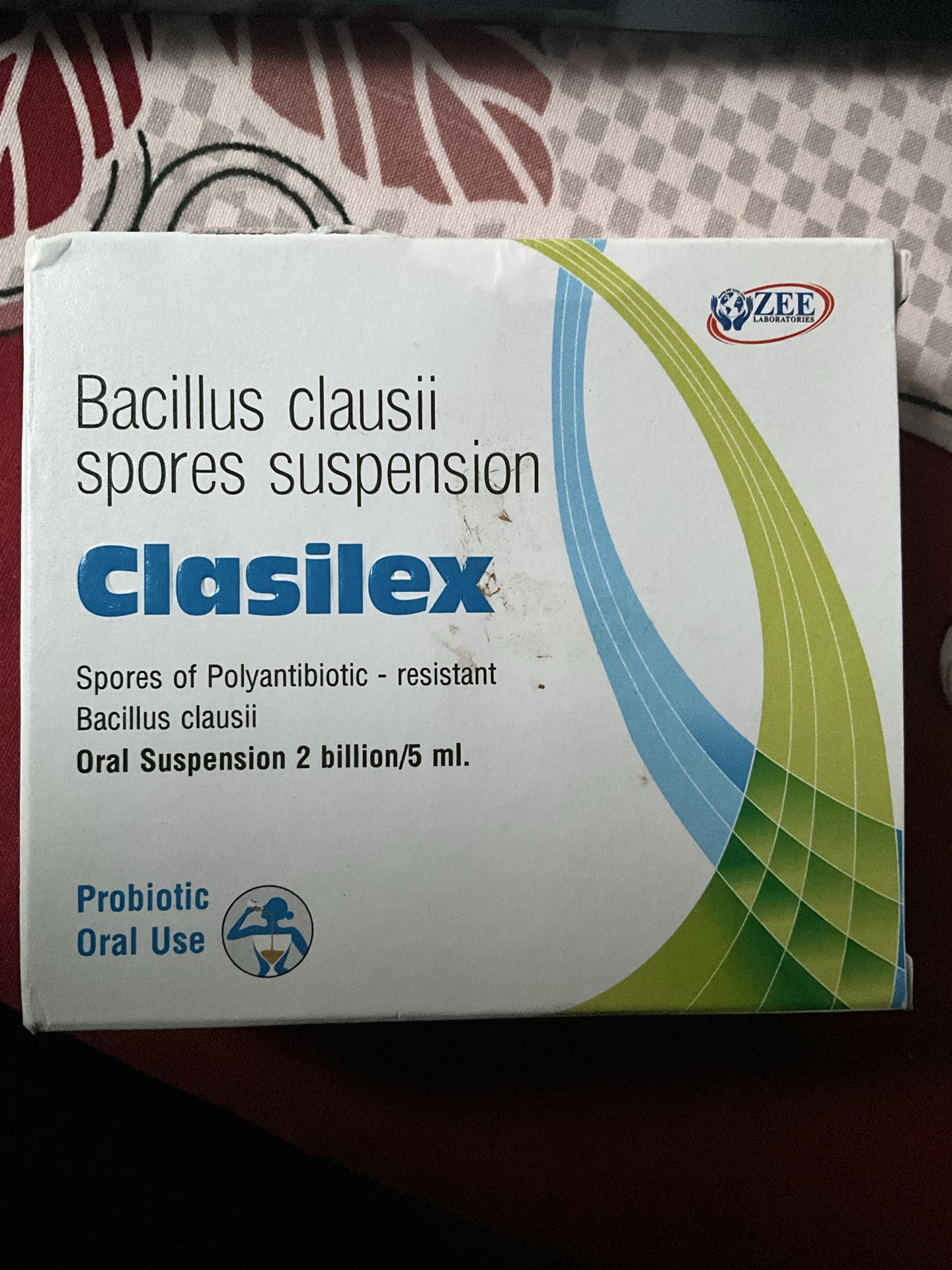
Clasilex is a specialized probiotic supplement containing Bacillus clausii spores suspension, designed to support digestive health and restore beneficial gut bacteria. This antibiotic-resistant probiotic offers unique advantages for maintaining intestinal balance, especially during and after antibiotic treatment.
What is Clasilex? Understanding Bacillus Clausii
Clasilex oral suspension contains 2 billion spores of Bacillus clausii per 5ml dose. Unlike traditional probiotics, Bacillus clausii is a spore-forming probiotic that can survive harsh stomach acid and reach the intestines where it’s needed most.


Key Features of Clasilex:
- Polyanitibiotic-resistant Bacillus clausii
- 2 billion spores per 5ml dose
- Oral suspension formula for easy administration
- Manufactured by ZEE Pharmaceuticals
- Suitable for both adults and children
The Science Behind Bacillus Clausii Probiotics
Bacillus clausii is a gram-positive, spore-forming bacterium that offers several advantages over traditional probiotics:
Antibiotic Resistance Benefits
The polyanitibiotic-resistant nature of Bacillus clausii means it can:
- Survive during antibiotic treatment
- Help prevent antibiotic-associated diarrhea
- Maintain gut flora balance when other probiotics cannot
- Support recovery after antibiotic courses
Spore Formation Advantages


Probiotic spores provide superior stability:
- Heat resistance for better storage
- Acid resistance for stomach survival
- Extended shelf life without refrigeration
- Consistent potency delivery
Health Benefits of Clasilex Probiotic Suspension
Digestive Health Support
- Restores gut microbiome balance
- Reduces symptoms of digestive disorders
- Supports healthy bowel movements
- Alleviates bloating and gas
Immune System Enhancement
- Strengthens intestinal barrier function
- Supports immune response regulation
- Helps prevent harmful bacteria overgrowth
- Promotes overall gut health
Special Applications
- Post-antibiotic recovery
- Management of acute diarrhea
- Support during intestinal infections
- Maintenance of digestive wellness


Dosage and Administration Guidelines
Standard Dosing for Clasilex:
- Adults: 5-10ml twice daily
- Children: 2.5-5ml twice daily
- Infants: As directed by healthcare provider
Best Practices:
- Take on an empty stomach when possible
- Can be mixed with water or juice
- Continue for 7-14 days or as recommended
- Safe for long-term use under medical supervision
Who Should Consider Clasilex Probiotics?
Ideal Candidates:
- Patients on antibiotic therapy
- Individuals with digestive imbalances
- Those experiencing traveler’s diarrhea
- People with compromised gut health
- Anyone seeking preventive digestive care
Special Populations:
- Elderly patients with digestive concerns
- Children with frequent stomach issues
- Immunocompromised individuals
- Patients with inflammatory bowel conditions
Clasilex vs. Other Probiotics: What Makes It Different?
Unique Advantages:
- Spore-based formulation survives stomach acid
- Antibiotic resistance allows concurrent use
- No refrigeration required for storage
- Clinically proven Bacillus clausii strain
- Liquid suspension for better absorption
Comparison with Traditional Probiotics:
- Higher survival rate through digestive tract
- More stable during storage and transport
- Effective during antibiotic treatment
- Better for acute digestive issues
Safety Profile and Side Effects
Generally Well Tolerated:
Clasilex probiotic has an excellent safety profile with minimal side effects:
- Rare mild digestive discomfort
- Occasional temporary bloating
- No significant drug interactions
- Safe for most age groups
Precautions:
- Consult healthcare provider before use
- Monitor for allergic reactions
- Adjust dosage based on response
- Inform doctor of all medications
Storage and Handling Instructions
Proper Storage:
- Store at room temperature
- Keep away from direct sunlight
- Ensure cap is tightly closed
- Check expiration date regularly
- No refrigeration necessary
Clinical Evidence Supporting Bacillus Clausii
Research studies have demonstrated the effectiveness of Bacillus clausii probiotics in:
- Reducing antibiotic-associated diarrhea
- Supporting immune function
- Maintaining intestinal barrier integrity
- Preventing harmful bacterial overgrowth
When to Consult Healthcare Providers
Seek Medical Advice If:
- Symptoms persist beyond expected timeframe
- Severe digestive symptoms occur
- Allergic reactions develop
- Concurrent medical conditions exist
- Taking multiple medications
Frequently Asked Questions About Clasilex
Can I take Clasilex with antibiotics?
Yes, the antibiotic-resistant nature of Bacillus clausii allows safe concurrent use.
How long should I take Clasilex?
Typical courses range from 7-14 days, but long-term use may be beneficial for some individuals.
Is Clasilex suitable for children?
Yes, with appropriate dosage adjustments for age and weight.
Conclusion: Choosing Clasilex for Digestive Health
Clasilex Bacillus clausii spores suspension represents an advanced approach to probiotic supplementation. Its unique antibiotic-resistant properties and spore-based formulation make it an excellent choice for maintaining digestive health, especially during challenging times when traditional probiotics may not be effective.
Whether you’re dealing with antibiotic-associated digestive issues, seeking preventive gut health support, or managing acute digestive symptoms, Clasilex offers a scientifically-backed solution that can help restore and maintain your intestinal balance.
Key Takeaways:
- Bacillus clausii is uniquely resistant to antibiotics
- Spore-based probiotics offer superior survival rates
- Clasilex suspension is easy to administer
- Suitable for various age groups with proper dosing
- Excellent safety profile with proven benefits
Disclaimer: This information is for educational purposes only and should not replace professional medical advice. Always consult with a healthcare provider before starting any new supplement regimen, especially if you have existing health conditions or are taking medications.
Keywords: Clasilex, Bacillus clausii, probiotic spores, antibiotic-resistant probiotics, oral suspension, digestive health, gut microbiome, probiotic benefits, ZEE Pharmaceuticals, polyanitibiotic-resistant