Monthly Archives: September 2026

Bromadiolone toxicokinetics: diagnosis and treatment implications

Bromadiolone Poisoning Treatment: Vitamin K1, INR Monitoring and Emergency Management

Bromadiolone poisoning is an important cause of potentially severe and prolonged bleeding because bromadiolone is a long-acting anticoagulant rodenticide (LAAR), commonly called a superwarfarin. It inhibits vitamin K recycling and consequently reduces the activity of vitamin K–dependent coagulation factors II, VII, IX and X.

Unlike ordinary warfarin exposure, bromadiolone poisoning may produce prolonged coagulopathy lasting weeks to months, and patients may require prolonged treatment with vitamin K1 (phytomenadione) and repeated INR monitoring.

Clinical pearl: A patient can appear clinically well initially and subsequently develop severe coagulopathy. A normal INR early after exposure does not necessarily exclude clinically important toxicity.


What is Bromadiolone?

Bromadiolone is a second-generation 4-hydroxycoumarin anticoagulant rodenticide. It belongs to the long-acting anticoagulant rodenticide group along with compounds such as brodifacoum and difenacoum.

It is considerably more persistent than conventional warfarin and can produce prolonged suppression of vitamin K–dependent coagulation.

Mechanism of bromadiolone poisoning

Bromadiolone inhibits vitamin K epoxide reductase, preventing regeneration of active vitamin K.

This results in impaired γ-carboxylation and activation of:

  • Factor II
  • Factor VII
  • Factor IX
  • Factor X
  • Protein C
  • Protein S

The result is progressive impairment of coagulation and an increased risk of spontaneous or trauma-related bleeding.


Clinical Features of Bromadiolone Poisoning

Patients may initially have no symptoms.

As coagulopathy develops, manifestations may include:

  • Epistaxis
  • Gingival bleeding
  • Easy bruising
  • Hematuria
  • Hematemesis
  • Melena
  • Hematochezia
  • Menorrhagia
  • Intramuscular bleeding
  • Hemarthrosis
  • Retroperitoneal bleeding
  • Intracranial hemorrhage
  • Hemoperitoneum
  • Severe anemia
  • Hemorrhagic shock

Intracranial hemorrhage is an uncommon but potentially life-threatening complication.


Initial Assessment

Management begins with stabilization and assessment of bleeding risk.

ABCDE assessment

A — Airway

Assess airway protection, particularly in patients with:

  • Altered consciousness
  • Intracranial hemorrhage
  • Massive upper gastrointestinal bleeding

B — Breathing

Assess:

  • Respiratory rate
  • Oxygen saturation
  • Respiratory distress

C — Circulation

Assess:

  • Heart rate
  • Blood pressure
  • Peripheral perfusion
  • Capillary refill
  • Evidence of active bleeding
  • Shock

Establish IV access and obtain blood samples early.

D — Disability

Assess:

  • Glasgow Coma Scale
  • Pupils
  • Focal neurological deficits
  • Headache
  • Seizures

Consider intracranial hemorrhage when neurological symptoms occur in a patient with severe coagulopathy.

E — Exposure

Look carefully for:

  • Bruising
  • Petechiae
  • Hematomas
  • Hematuria
  • Gastrointestinal bleeding
  • Injection or trauma sites

Investigations in Bromadiolone Poisoning

Important investigations include:

Essential tests

  • PT/INR
  • aPTT
  • CBC with platelet count
  • Hemoglobin/hematocrit
  • Blood group and crossmatch
  • Fibrinogen
  • Renal function
  • Liver function tests
  • Serum electrolytes

Additional tests when indicated

  • Factor II, VII, IX and X levels
  • Mixing study
  • Specific bromadiolone/superwarfarin assay
  • CT brain for neurological symptoms
  • Ultrasound/CT for suspected internal bleeding

Bromadiolone poisoning characteristically produces a vitamin K–dependent factor deficiency pattern, particularly involving factors II, VII, IX and X.


Bromadiolone Poisoning Treatment

The two major principles of treatment are:

  1. Replace vitamin K1 to overcome the anticoagulant effect
  2. Rapidly replace deficient coagulation factors when there is significant or life-threatening bleeding

1. Vitamin K1 — The Specific Antidote

Vitamin K1 (phytomenadione/phytonadione) is the cornerstone of treatment.

It restores the availability of reduced vitamin K and allows synthesis of functional vitamin K–dependent clotting factors.

Importantly, vitamin K does not immediately replace circulating clotting factors. Therefore, a patient with life-threatening hemorrhage may require coagulation-factor replacement in addition to vitamin K.

Route of vitamin K1

Depending on severity:

  • Oral vitamin K1 — useful for stable patients and prolonged treatment
  • IV vitamin K1 — preferred when rapid treatment is required or oral administration is not feasible

IV vitamin K should be administered cautiously because serious hypersensitivity/anaphylactoid reactions have been reported.


Vitamin K1 Dose in Bromadiolone Poisoning

There is no universally standardized dose or duration for bromadiolone poisoning.

The required dose depends on:

  • INR
  • Severity of coagulopathy
  • Presence or absence of bleeding
  • Amount/type of rodenticide exposure
  • Response to treatment
  • Recurrence of coagulopathy after dose reduction or interruption

Published cases have used a wide range of regimens, including repeated IV vitamin K followed by high-dose oral vitamin K.

For severe superwarfarin poisoning, high-dose vitamin K1 administered repeatedly throughout the day may be necessary, with dosing adjusted according to the INR and clinical response. Some toxicology literature reports doses as high as 50–100 mg/day or more in severe cases, but these doses should be individualized with toxicology/hematology guidance rather than applied as a routine dose.

Important

Do not use a single fixed vitamin K dose for every bromadiolone ingestion.

The appropriate regimen should be guided by:

Clinical bleeding + INR/PT response + toxicology consultation


2. Management of Active or Life-Threatening Bleeding

Vitamin K alone may be insufficient initially because the liver requires time to synthesize new coagulation factors.

For patients with major, life-threatening bleeding, rapid coagulation-factor replacement is required.

Options include:

4-factor PCC

4-factor prothrombin complex concentrate (PCC) provides factors:

  • II
  • VII
  • IX
  • X

and can rapidly correct severe vitamin K antagonist–associated coagulopathy.

Fresh Frozen Plasma

FFP can also replace deficient coagulation factors.

It may be used when PCC is unavailable or according to local protocol.

Packed RBCs

Packed red blood cells should be administered when significant blood loss has caused clinically important anemia or hemorrhagic shock.

The choice and dose of blood products should be individualized according to bleeding severity, hemoglobin, hemodynamic status and local transfusion protocols.

Key principle

Major bleeding = vitamin K1 + rapid coagulation-factor replacement + definitive control of the bleeding source.


3. Control the Source of Bleeding

Correction of coagulopathy is not enough if active bleeding continues.

Depending on the site, management may include:

  • Endoscopic hemostasis
  • Surgical intervention
  • Interventional radiology
  • Neurosurgical intervention
  • Local pressure
  • Gynecological intervention
  • Management of gastrointestinal bleeding

A published case of severe bromadiolone-associated intracranial/other hemorrhage illustrates the need for both correction of coagulopathy and definitive treatment of the bleeding source.


4. Activated Charcoal and Gastric Decontamination

Routine gastrointestinal decontamination is not the main treatment for established bromadiolone poisoning.

Evidence summarized in toxicology reviews indicates that multiple-dose activated charcoal has not demonstrated reliable benefit for superwarfarin poisoning.

Any consideration of activated charcoal after a recent ingestion should therefore be individualized according to:

  • Time since ingestion
  • Amount ingested
  • Airway protection
  • Toxicology advice
  • Product formulation

Do not delay resuscitation or treatment of hemorrhage for gastrointestinal decontamination.


5. INR Monitoring

INR is the most practical laboratory marker for monitoring the anticoagulant effect.

A patient with significant exposure should have serial:

  • PT/INR
  • aPTT
  • CBC
  • Hemoglobin

The frequency depends on the severity of poisoning and treatment response.

In severe poisoning, INR may need to be checked frequently during initial stabilization, followed by less frequent monitoring once a stable vitamin K regimen has been established.


Why Long-Term Treatment May Be Necessary

This is one of the most important differences between bromadiolone and ordinary warfarin poisoning.

Bromadiolone is highly lipophilic and has prolonged persistence in the body. Human pharmacokinetic data demonstrate a prolonged terminal elimination phase; one clinical study estimated a terminal half-life of approximately 24 days.

Therefore:

Stopping vitamin K too early → recurrent INR elevation → recurrent bleeding.

Cases have documented recurrence of severe coagulopathy after vitamin K was discontinued despite an initially normal INR.


When Can Vitamin K Be Stopped?

There is no universally accepted evidence-based stopping rule for bromadiolone poisoning.

A normal INR while the patient is receiving vitamin K does not necessarily mean that bromadiolone has been eliminated.

A commonly used approach is:

  1. Stabilize the INR with vitamin K.
  2. Continue vitamin K while the toxic effect persists.
  3. Gradually reduce/taper treatment when appropriate.
  4. Stop vitamin K under specialist supervision.
  5. Recheck PT/INR approximately 48–72 hours after stopping vitamin K.
  6. Restart treatment if significant coagulopathy recurs.

Where available, quantitative serum bromadiolone/superwarfarin testing can provide additional information, although there is no universally validated concentration threshold that independently determines when treatment can safely stop.


Bromadiolone Poisoning: Practical Treatment Algorithm

Suspected bromadiolone ingestion

Assess ABCDE + bleeding

Obtain PT/INR ± aPTT, CBC and other baseline investigations

Is there major/life-threatening bleeding?

YES

→ Vitamin K1
→ 4-factor PCC or FFP for rapid factor replacement
→ RBC transfusion when indicated
→ Definitive control of bleeding
→ Serial INR monitoring
→ Toxicology/hematology consultation

NO

→ Assess exposure and coagulation profile
→ Serial PT/INR monitoring
→ Vitamin K1 when clinically indicated
→ Continue monitoring because delayed/prolonged coagulopathy can occur

INR controlled?

YES

→ Continue appropriately dosed vitamin K1
→ Regular INR monitoring
→ Gradual reduction when appropriate

NO

→ Check adherence
→ Reassess ongoing exposure/re-exposure
→ Increase/adjust vitamin K under specialist guidance
→ Evaluate for ongoing bleeding

Considering stopping vitamin K?

→ Stop only after adequate clinical assessment
→ Recheck INR after approximately 48–72 hours
→ Restart treatment if coagulopathy recurs


Bromadiolone Poisoning in Children

Children may accidentally ingest rodenticide bait.

A child who has ingested a small amount may remain asymptomatic, but the risk assessment depends on:

  • Exact product
  • Active ingredient
  • Concentration
  • Amount ingested
  • Child’s weight
  • Time since ingestion
  • Repeated versus single exposure

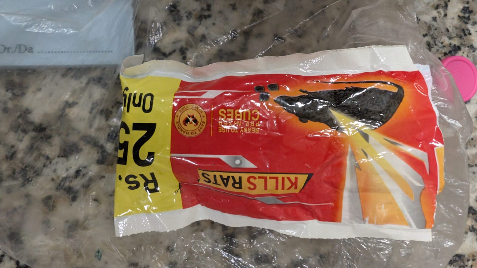
Do not automatically assume that every rodenticide product contains bromadiolone. The product label should be checked whenever possible.

For children with suspected anticoagulant rodenticide exposure, consultation with a poison center or medical toxicologist is recommended.


Important Differential Diagnoses

A markedly prolonged PT/INR should not automatically be attributed to bromadiolone.

Consider:

  • Warfarin exposure
  • Other anticoagulant rodenticides
  • Vitamin K deficiency
  • Liver disease
  • Disseminated intravascular coagulation
  • Acquired factor inhibitors
  • Congenital coagulation-factor deficiencies
  • Malabsorption
  • Drug interactions

Superwarfarin poisoning should particularly be considered in a patient with unexplained prolonged PT/INR and bleeding, especially when the history is unclear.


Key Differences: Warfarin vs Bromadiolone

FeatureWarfarinBromadiolone
UseTherapeutic anticoagulantRodenticide
ClassVitamin K antagonistLong-acting vitamin K antagonist
Common nameWarfarinSuperwarfarin
Duration of effectUsually shorterProlonged
CoagulopathyUsually manageable over daysMay persist weeks–months
AntidoteVitamin K1Vitamin K1
Severe bleedingFactor replacement may be requiredFactor replacement may be required
Long-term vitamin KSometimesFrequently required in severe poisoning
MonitoringINRSerial INR/PT

Important Clinical Pearls

1. A normal early INR does not completely exclude toxicity.

The anticoagulant effect can be delayed because existing circulating clotting factors must first decline.

2. Vitamin K1 is the specific treatment.

Bromadiolone poisoning is fundamentally a vitamin K antagonist poisoning.

3. Vitamin K does not immediately correct major hemorrhage.

In life-threatening bleeding, rapidly replace coagulation factors with 4-factor PCC or FFP, according to availability and local protocol.

4. Treatment may last for months.

Severe bromadiolone poisoning can require prolonged vitamin K1 therapy because of its long persistence.

5. Do not stop vitamin K simply because the INR becomes normal.

The INR may normalize because of administered vitamin K while the rodenticide remains in the body.

6. Monitor after stopping therapy.

A recurrence of INR elevation after vitamin K withdrawal strongly suggests persistent anticoagulant activity.

7. Always identify the active ingredient.

“Rat poison” is not a diagnosis. Different rodenticides have different toxic mechanisms and treatments.


Summary

Bromadiolone poisoning is a potentially life-threatening superwarfarin poisoning characterized by prolonged vitamin K–dependent coagulopathy.

The cornerstone of treatment is:

Vitamin K1 + INR-guided monitoring + rapid factor replacement when significant bleeding is present.

Patients with severe poisoning may require high-dose and prolonged vitamin K1 therapy for weeks or months. Treatment should not be discontinued solely because the INR has normalized while the patient is receiving vitamin K. After vitamin K is eventually stopped, repeat coagulation testing after approximately 48–72 hours can help identify recurrent anticoagulation.

Because bromadiolone poisoning can be prolonged and potentially fatal, significant exposures, abnormal coagulation studies, or any active bleeding warrant urgent medical evaluation and specialist toxicology/hematology input.


Frequently Asked Questions

What is the antidote for bromadiolone poisoning?

Vitamin K1 (phytomenadione/phytonadione) is the specific antidote for bromadiolone-induced vitamin K antagonism.

How long does bromadiolone poisoning last?

Severe poisoning can persist for weeks to months, reflecting the prolonged persistence of bromadiolone and its anticoagulant effect.

Does bromadiolone poisoning cause bleeding?

Yes. Severe poisoning can cause epistaxis, gum bleeding, hematuria, gastrointestinal bleeding, internal hemorrhage and, rarely, intracranial hemorrhage.

Can bromadiolone poisoning be treated?

Yes. Early recognition, vitamin K1 therapy, appropriate coagulation-factor replacement and close monitoring can successfully reverse the coagulopathy.

Is vitamin K enough for severe bleeding?

Not necessarily. Vitamin K restores coagulation factor production but does not provide an immediate supply of functional factors. Patients with life-threatening bleeding may require 4-factor PCC or FFP in addition to vitamin K1.

Can bromadiolone poisoning recur after treatment?

Yes. Recurrent coagulopathy can occur after vitamin K is stopped because bromadiolone may remain active in the body for a prolonged period.


SEO Keywords

Primary keyword:
Bromadiolone poisoning treatment

Secondary keywords:

  • Bromadiolone poisoning
  • Bromadiolone treatment
  • Bromadiolone antidote
  • Vitamin K for bromadiolone poisoning
  • Bromadiolone toxicity
  • Bromadiolone overdose
  • Bromadiolone rodenticide poisoning
  • Superwarfarin poisoning treatment
  • Anticoagulant rodenticide poisoning
  • Vitamin K1 treatment
  • Bromadiolone INR
  • Bromadiolone coagulopathy
  • Rat poison poisoning treatment
  • Rodenticide poisoning treatment
  • Bromadiolone bleeding
  • Bromadiolone poisoning management

Direct Coombs Test (DCT): Indications, Clinical Uses, and Interpretation

The Direct Coombs Test (DCT), also known as the Direct Antiglobulin Test (DAT), is an important laboratory investigation used to detect immunoglobulins or complement components attached to the surface of red blood cells (RBCs). It is particularly useful in the evaluation of suspected immune-mediated hemolysis, autoimmune hemolytic anemia, hemolytic disease of the fetus and newborn, and hemolytic transfusion reactions.

This article explains the principle of the Direct Coombs Test, the major clinical conditions in which it is used, and how to interpret its results in pediatric and neonatal practice.

What Is the Direct Coombs Test?

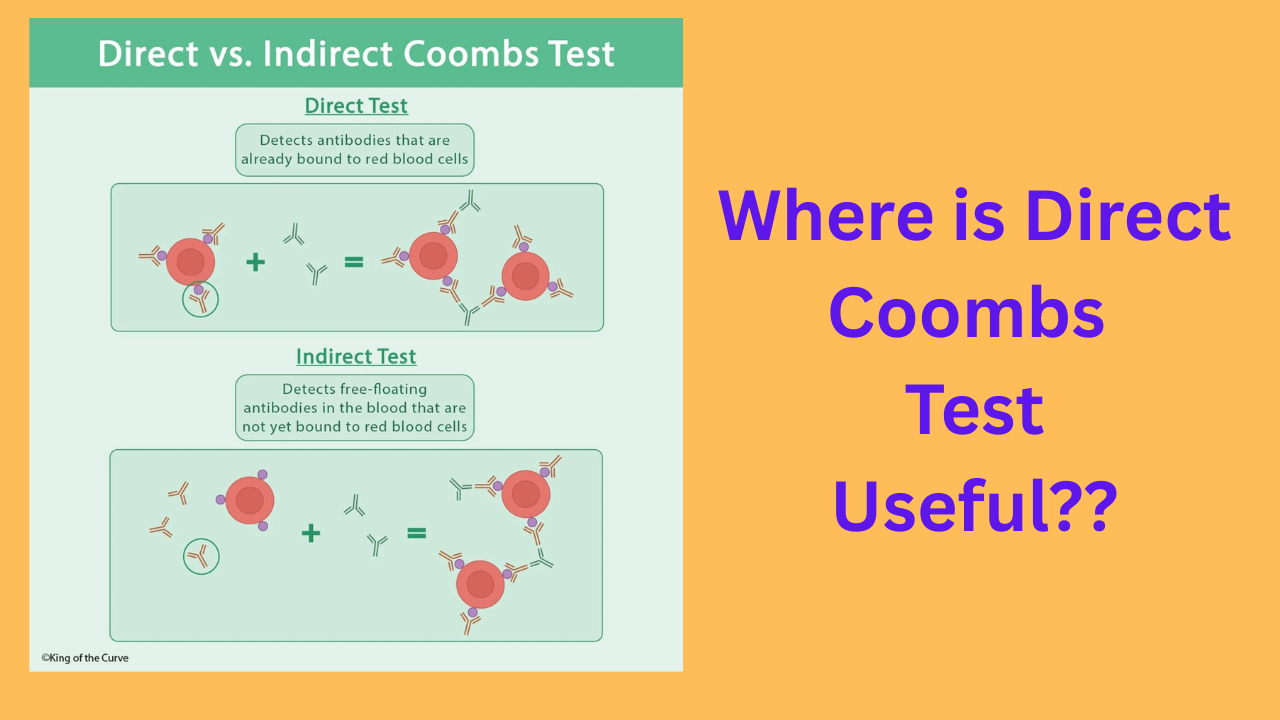
The Direct Coombs Test detects IgG antibodies and/or complement components attached to the patient’s red blood cells in vivo. These substances may be responsible for immune-mediated destruction of RBCs.

In the laboratory, the patient’s RBCs are washed to remove unbound proteins. Antihuman globulin reagent is then added. If immunoglobulin or complement is attached to the RBC surface, the reagent may cause visible agglutination, resulting in a positive test.

Principle of the Direct Antiglobulin Test

  1. Red blood cells are collected from the patient.
  2. The RBCs are washed to remove unbound antibodies and plasma proteins.
  3. Antihuman globulin reagent is added.
  4. Agglutination indicates that immunoglobulin and/or complement is attached to the RBCs.

A positive DCT indicates that RBCs are coated with immunoglobulin or complement. However, it does not independently establish that clinically significant hemolysis is occurring.

Conditions in Which the Direct Coombs Test Is Used

The DCT is primarily used when immune-mediated red blood cell destruction is suspected. The following are the major clinical conditions associated with its use.

1. Autoimmune Hemolytic Anemia

Autoimmune hemolytic anemia (AIHA) occurs when the immune system produces antibodies against the patient’s own red blood cells, resulting in premature RBC destruction.

The Direct Coombs Test is an important investigation in suspected AIHA because it can demonstrate immunoglobulin or complement attached to the RBC surface.

  • Warm autoimmune hemolytic anemia: Usually associated with IgG coating of RBCs. The DCT is commonly positive for IgG, with or without complement.
  • Cold agglutinin disease: Commonly associated with complement, particularly C3, attached to RBCs. The IgG component may be negative.

Clinical findings that may support a diagnosis of AIHA include pallor, jaundice, dark urine, splenomegaly, anemia, reticulocytosis, elevated lactate dehydrogenase, and reduced haptoglobin.

2. Hemolytic Disease of the Fetus and Newborn

Hemolytic disease of the fetus and newborn (HDFN) occurs when maternal IgG antibodies cross the placenta and bind to antigens on fetal or neonatal red blood cells.

The DCT is used on the newborn’s RBCs to detect maternal antibodies attached to the cells.

It is particularly relevant in cases involving:

  • Rh incompatibility, including maternal anti-D antibodies.
  • ABO incompatibility, especially when maternal anti-A or anti-B IgG antibodies affect the newborn.
  • Other clinically significant maternal red-cell alloantibodies, such as anti-c or anti-K.

A newborn with jaundice and anemia, especially in the presence of maternal–fetal blood-group incompatibility, may require a DCT as part of the evaluation.

Important neonatal point: A positive DCT does not necessarily mean that the newborn has clinically significant hemolysis. The result should be interpreted together with hemoglobin, bilirubin, reticulocyte count, peripheral blood smear, and the clinical condition of the baby.

3. ABO Incompatibility in Newborns

ABO incompatibility can occur when a mother has blood group O and the newborn has blood group A or B. Maternal IgG anti-A or anti-B antibodies may cross the placenta and bind to neonatal RBCs.

The DCT may be positive in an affected newborn. However, the test can be negative in some cases of clinically significant ABO hemolysis, and a positive result does not always indicate substantial hemolysis.

Therefore, the DCT should not be used in isolation to determine the severity of neonatal jaundice or to decide on treatment.

4. Rh Isoimmunization

Rh isoimmunization occurs when a mother develops antibodies against Rh antigens present on fetal red blood cells. Maternal anti-D IgG is a classic example.

When these antibodies cross the placenta, they may cause fetal or neonatal hemolysis. The Direct Coombs Test can detect maternal antibodies attached to the newborn’s RBCs.

Depending on the severity, affected newborns may develop anemia, jaundice, or, in severe cases, hydrops fetalis.

5. Hemolytic Transfusion Reactions

The Direct Coombs Test may be useful in the investigation of suspected immune-mediated hemolytic transfusion reactions.

Acute Hemolytic Transfusion Reaction

An acute hemolytic transfusion reaction may occur when incompatible blood is transfused, leading to immune-mediated destruction of transfused RBCs. The DCT may become positive when immunoglobulin or complement is attached to RBCs.

Delayed Hemolytic Transfusion Reaction

A delayed hemolytic transfusion reaction usually occurs days to weeks after a transfusion when an alloantibody-mediated immune response leads to destruction of transfused RBCs.

The DCT may be positive during the investigation, although the result depends on the antibody, the timing of testing, and the degree of RBC coating.

Evaluation should also include transfusion history, antibody screening, antibody identification, hemoglobin, bilirubin, reticulocyte count, and other appropriate hemolysis investigations.

6. Drug-Induced Immune Hemolytic Anemia

Some medications can trigger immune-mediated hemolytic anemia. Drug-induced immune hemolytic anemia may occur through different mechanisms, including drug-dependent or drug-independent antibodies.

The DCT may be positive for IgG, complement, or both, depending on the mechanism involved.

When drug-induced hemolysis is suspected, the medication history is essential. The laboratory findings must be interpreted with clinical evidence of hemolysis and, when necessary, specialized immunohematological testing.

7. Systemic Lupus Erythematosus

Systemic lupus erythematosus (SLE) and other autoimmune disorders may be associated with autoimmune hemolytic anemia.

The DCT can be positive when RBC-bound antibodies or complement are present. However, a positive DCT in a patient with SLE does not necessarily indicate active hemolysis.

The diagnosis of autoimmune hemolytic anemia requires correlation with anemia, evidence of increased RBC destruction, and exclusion of other causes.

8. Lymphoproliferative Disorders

Lymphoproliferative disorders, including certain leukemias and lymphomas, may be associated with autoimmune hemolytic anemia.

The DCT can help identify RBC-bound immunoglobulin or complement in patients with suspected immune-mediated hemolysis. The result should be interpreted alongside the patient’s clinical presentation and hematological investigations.

Direct Coombs Test: DCT vs. Indirect Coombs Test

The Direct and Indirect Coombs Tests are both antiglobulin tests, but they detect different targets.

Feature Direct Coombs Test (DCT) Indirect Coombs Test (ICT)
Also called Direct Antiglobulin Test (DAT) Indirect Antiglobulin Test (IAT)
What it detects IgG and/or complement attached to RBCs Antibodies present in serum or plasma that can bind to RBC antigens
Where the antibodies are detected On the RBC surface In the serum or plasma
Major clinical use Immune hemolysis and HDFN evaluation Pretransfusion antibody screening and antenatal antibody screening
Neonatal application Detects maternal antibodies attached to neonatal RBCs Detects clinically significant antibodies in maternal serum

How to Interpret a Positive DCT

A positive DCT means that immunoglobulin and/or complement has been detected on the surface of the tested RBCs.

A positive result may be seen in:

  • Autoimmune hemolytic anemia.
  • Hemolytic disease of the fetus and newborn.
  • Immune-mediated hemolytic transfusion reactions.
  • Drug-induced immune hemolytic anemia.
  • Some autoimmune and lymphoproliferative disorders.
  • Some patients without clinically important hemolysis.

A positive DCT is not synonymous with hemolytic anemia. The presence of RBC-bound immunoglobulin or complement must be interpreted in the context of the patient’s clinical findings and laboratory evidence.

Investigations to Correlate With DCT Results

When immune hemolysis is suspected, relevant investigations may include:

  • Complete blood count and hemoglobin concentration.
  • Reticulocyte count.
  • Total and indirect bilirubin.
  • Lactate dehydrogenase (LDH).
  • Haptoglobin, when appropriate.
  • Peripheral blood smear.
  • Blood group and antibody screening.
  • Clinical history of transfusion, medication exposure, and maternal–fetal blood-group incompatibility.

Can the Direct Coombs Test Be Negative in Hemolysis?

Yes. A negative DCT does not completely exclude immune-mediated hemolysis.

Possible explanations include:

  • The quantity of RBC-bound antibody is below the detection threshold of the test.
  • The antibody class or subtype is not adequately detected by the reagent used.
  • The RBC-bound immunoglobulin has been removed or reduced before testing.
  • The hemolysis is nonimmune in origin.

In patients with strong clinical evidence of autoimmune hemolytic anemia but a negative routine DCT, further evaluation with an appropriate laboratory may be considered.

High-Yield Clinical Summary

  • DCT/DAT: Detects IgG and/or complement attached to the patient’s RBCs.
  • Warm AIHA: Usually associated with IgG-positive RBCs.
  • Cold agglutinin disease: Commonly associated with C3-positive RBCs.
  • HDFN: DCT may detect maternal IgG attached to neonatal RBCs.
  • Transfusion reactions: DCT may support the evaluation of immune-mediated hemolysis.
  • Positive DCT: Does not automatically prove active hemolysis.
  • Negative DCT: Does not completely exclude immune hemolysis.
  • Clinical correlation: Always interpret the result alongside evidence of hemolysis and the clinical context.

Frequently Asked Questions (FAQs)

1. What is the main purpose of the Direct Coombs Test?

The main purpose of the DCT is to detect immunoglobulin or complement attached to the surface of red blood cells. It is commonly used in the evaluation of autoimmune hemolytic anemia, hemolytic disease of the fetus and newborn, and immune-mediated transfusion reactions.

2. Is the Direct Coombs Test positive in ABO incompatibility?

It may be positive when maternal IgG anti-A or anti-B antibodies bind to neonatal RBCs. However, not every newborn with ABO incompatibility has a positive DCT, and a positive result does not necessarily indicate significant hemolysis.

3. What is the difference between DCT and ICT?

The DCT detects antibodies or complement attached to RBCs, whereas the ICT detects antibodies in serum or plasma that can bind to RBC antigens. The DCT is commonly used in evaluating immune hemolysis, while the ICT is used in antibody screening and compatibility testing.

4. Does a positive DCT confirm autoimmune hemolytic anemia?

No. A positive DCT supports the presence of RBC-bound immunoglobulin or complement, but autoimmune hemolytic anemia requires evidence of hemolysis and appropriate clinical correlation.

5. Can the DCT be negative in autoimmune hemolytic anemia?

Yes. Some patients with autoimmune hemolytic anemia may have a negative routine DCT because of low levels of RBC-bound antibodies or limitations of the testing method.

Conclusion

The Direct Coombs Test (DCT) is an essential investigation for detecting immunoglobulin or complement attached to red blood cells. Its major clinical applications include autoimmune hemolytic anemia, hemolytic disease of the fetus and newborn, immune-mediated transfusion reactions, and drug-induced immune hemolysis.

For pediatricians and neonatologists, the DCT is particularly important in evaluating newborns with suspected immune-mediated hemolysis. Nevertheless, the test result should always be interpreted together with the clinical condition, hemoglobin, bilirubin, reticulocyte count, and other relevant investigations.

Educational note: This article is intended for medical education. Diagnosis and treatment should be guided by the clinical context, institutional protocols, and appropriate specialist consultation.

Neutropenia in Children: Definition, ANC Classification, Causes and Management

1. Definition of neutropenia

Neutropenia = ANC below the age-appropriate normal lower limit.

In older children/adults, a commonly used cutoff is:

ANC <1,500 cells/µL (1.5 × 10⁹/L)

However, ANC must be interpreted according to age and population, particularly in infants and young children. The infection risk correlates much more strongly with the severity and duration of neutropenia than simply with whether ANC is below 1,500. (MSD Manuals)

For pediatrics, the key is to distinguish neutropenia (a laboratory finding) from febrile neutropenia (a clinical emergency defined by fever + sufficiently low ANC).

Calculate ANC

ANC=WBC×(%neutrophils+%bands)100\boxed{ANC = WBC \times \frac{(\%neutrophils+\%bands)}{100}}

Example:

WBC = 2,000/µL
Neutrophils = 20%
Bands = 5%

ANC = 2,000 × 25/100 = 500/µL


2. Classification by ANC

ANCClassificationInfection risk
1,000–1,500/µLMildUsually minimal
500–1,000/µLModerateIncreased
<500/µLSevereHigh
<200/µLProfoundVery high; inflammatory response may be minimal

These are the conventional severity categories used clinically. (MSD Manuals)

Important threshold

ANC <500/µL = severe neutropenia.

At this level, organisms from the patient’s own oral/GI flora can produce serious infection. At ANC <200/µL, signs of inflammation can be particularly muted. (MSD Manuals)


3. Classification by duration

Acute neutropenia

Develops over hours to days.

Common causes:

  • Viral infection
  • Drugs
  • Chemotherapy
  • Severe bacterial infection/sepsis
  • Acute marrow suppression

Chronic neutropenia

Persists for months or longer.

Examples:

  • Chronic benign/idiopathic neutropenia
  • Autoimmune neutropenia
  • Congenital neutropenia
  • Cyclic neutropenia
  • Bone marrow disorders

4. Classification by mechanism

A. Decreased production

  • Chemotherapy/radiation
  • Aplastic anemia
  • Leukemia/marrow infiltration
  • Myelodysplasia
  • B12/folate deficiency
  • Severe malnutrition
  • Congenital neutropenia

B. Increased destruction/consumption

  • Autoimmune neutropenia
  • Drug-induced immune neutropenia
  • Severe infections/sepsis

C. Sequestration

  • Hypersplenism

There can also be mixed mechanisms. (MSD Manuals)


5. What exactly is febrile neutropenia?

The classic oncology definition is:

Fever

Either:

  • Single temperature ≥38.3°C, OR
  • ≥38.0°C sustained for ≥1 hour

AND

Neutropenia

  • ANC <500/µL, OR
  • ANC expected to fall to <500/µL within the next 48 hours

(IDMP)

So:

Fever + ANC <500 = febrile neutropenia until proven otherwise.

This is particularly important in children receiving chemotherapy or hematopoietic stem-cell transplantation.


6. What is NOT necessarily called febrile neutropenia?

This is an important distinction.

Scenario 1 — Fever + ANC 800

Not conventionally called febrile neutropenia because ANC is >500.

But the child has:

Fever with moderate neutropenia

The clinical approach depends heavily on the underlying disease and immune status.


Scenario 2 — Fever + ANC 1,200

Not febrile neutropenia.

It’s simply:

Fever with mild neutropenia

Look for an underlying viral illness, drug effect, etc.


Scenario 3 — ANC <500 but NO fever

This is:

Severe/profound afebrile neutropenia

Not febrile neutropenia.

However, it may still require urgent assessment depending on the cause, duration and clinical condition.


Scenario 4 — Fever + transient viral neutropenia in an otherwise well child

For example:

4-year-old with viral URI
Temperature 39°C
ANC 700
Otherwise well

This is febrile illness with moderate neutropenia, not classic febrile neutropenia.

You shouldn’t automatically apply the chemotherapy febrile-neutropenia pathway simply because the child happens to have a low ANC.


7. The important pediatric exception

This is where clinicians sometimes get confused.

A child with:

fever + ANC <500

may technically fulfill the numerical definition of febrile neutropenia.

But management depends on the clinical context.

For example:

Child receiving chemotherapy

Fever + ANC <500
Treat as febrile neutropenia
→ urgent cultures + empiric antipseudomonal antibiotic.

Previously healthy child with viral illness

Fever + ANC <500
→ technically febrile severe neutropenia, but this is not automatically equivalent to chemotherapy-associated febrile neutropenia.

The risk profile is different.


8. Conditions where you should NOT casually label it “febrile neutropenia”

Think of these separately:

SituationBetter terminology
Viral infection + ANC 800 + feverFebrile illness with moderate neutropenia
Viral infection + ANC 300 but clinically wellSevere neutropenia associated with viral infection
Afebrile ANC 300Severe/profound neutropenia
Chronic benign neutropenia + feverFever in a child with chronic neutropenia; assess individually
Cyclic neutropenia + fever during neutropenic phaseFebrile episode during cyclic neutropenia
Autoimmune neutropenia + feverFever with autoimmune neutropenia
Chemotherapy + fever + ANC <500Classic febrile neutropenia
HSCT + fever + ANC <500Febrile neutropenia / high-risk febrile neutropenia

The distinction matters because chemotherapy-associated febrile neutropenia is a high-risk infectious syndrome, whereas many otherwise healthy children develop transient neutropenia during viral infections.


9. Management of neutropenia

A. First question: Is the child febrile or clinically unwell?

If fever + severe neutropenia

Treat as a potentially serious infection.

Immediate:

  1. ABC/clinical stability
  2. Full examination
  3. CBC + differential
  4. Blood cultures
  5. Urine evaluation/culture when appropriate
  6. Evaluate for focal infection
  7. Start appropriate empiric antibiotics promptly

For high-risk pediatric oncology patients, institutional protocols commonly use an antipseudomonal β-lactam such as cefepime, with escalation/additional coverage based on instability, focal infection, resistant organisms and local epidemiology. (IDMP)

Do not wait for the ANC to recover before treating a clinically significant suspected infection.


10. If the child is afebrile

Management depends on:

  • ANC
  • Duration of neutropenia
  • Age
  • Previous infections
  • Underlying disease
  • Chemotherapy/HSCT status
  • Medications
  • Clinical appearance

Mild ANC 1,000–1,500

Usually:

Observe + repeat CBC + investigate cause if persistent.

Moderate ANC 500–1,000

Look for cause and follow trend.

Severe ANC <500

Need much closer assessment, particularly if persistent.

Profound ANC <200

Highest concern for serious infection; clinical signs may be deceptively subtle. (MSD Manuals)


11. Treat the cause

Examples:

Drug-induced
→ stop suspected offending drug when appropriate.

Viral
→ usually supportive treatment and serial ANC.

Autoimmune neutropenia
→ often observation if child is clinically well; treatment depends on severity/infections.

Congenital/severe chronic neutropenia
→ hematology evaluation; G-CSF may be indicated.

Chemotherapy-induced
→ oncology-directed management ± G-CSF depending on regimen/risk.

Nutritional
→ correct B12/folate/nutritional deficiency.

Bone marrow failure/infiltration
→ urgent hematology evaluation.

G-CSF is particularly useful in selected severe/chronic neutropenia and in prevention/reduction of chemotherapy-related neutropenia. (MSD Manuals)


High-yield exam summary

Neutropenia = low ANC.

ANC:

  • 1000–1500 → mild
  • 500–1000 → moderate
  • <500 → severe
  • <200 → profound

Febrile neutropenia:

Fever ≥38.3°C once OR ≥38.0°C for ≥1 h + ANC <500/µL (or expected to fall <500 within 48 h).

And remember:

ANC <500 without fever = severe neutropenia, NOT febrile neutropenia.

Fever with ANC 500–1000 = febrile illness with moderate neutropenia, not the conventional febrile-neutropenia definition.

The classic “febrile neutropenia” pathway is particularly relevant to children receiving chemotherapy/HSCT; don’t automatically equate every viral-associated neutropenia in a healthy child with chemotherapy-associated FN. (IDMP)

Vitamin-Responsive Seizures in Children and Neonates: The Treatable Epilepsies You Should Not Miss

Vitamin-responsive seizures are uncommon, but they represent some of the most important treatable causes of neonatal and early-infantile epilepsy. Recognizing them early can prevent status epilepticus, developmental injury, and potentially irreversible neurological damage.

Introduction

Neonatal seizures are usually approached as an emergency, with hypoxic-ischemic injury, hypoglycemia, hypocalcemia, intracranial hemorrhage, stroke, infection, and structural brain abnormalities among the major considerations.

However, a small but particularly important group of seizures results from inborn errors of metabolism involving vitamins or vitamin-dependent enzymes. These seizures may be remarkably resistant to conventional antiseizure medications but respond dramatically to administration of the appropriate vitamin or cofactor.

The major clinically relevant disorders include:

  • Pyridoxine-dependent epilepsy (PDE)
  • Pyridoxal-5′-phosphate (PLP)-dependent epilepsy
  • Folinic acid-responsive seizures
  • Biotinidase deficiency
  • Biotin-thiamine-responsive basal ganglia disease
  • Other metabolic disorders in which vitamin/cofactor therapy can be seizure-modifying

Among these, vitamin B6-dependent epilepsies are particularly important in the neonate.


1. When should you suspect a vitamin-responsive seizure disorder?

The diagnosis should enter the differential particularly when a neonate or infant has:

Red flags

  1. Seizures beginning in the neonatal period without an obvious cause
  2. Frequent or recurrent seizures despite appropriate antiseizure medication
  3. Status epilepticus
  4. Abnormal EEG with burst suppression or marked discontinuity
  5. Seizures accompanied by unexplained encephalopathy
  6. Irritability, abnormal crying, vomiting, or feeding difficulty
  7. Unexplained metabolic acidosis or elevated lactate
  8. Seizures recurring after apparently successful treatment
  9. A previous sibling who died from unexplained neonatal seizures
  10. Consanguinity or a family history suggestive of an autosomal-recessive disorder
  11. Seizures with unusual movements, spasms, myoclonus, or autonomic features
  12. Seizures that respond dramatically to pyridoxine or PLP

The ILAE specifically recommends considering a trial of pyridoxine in neonates with clinical features suggestive of vitamin B6-dependent epilepsy and in neonates whose seizures remain unexplained and refractory to second-line antiseizure medication.


2. Pyridoxine-dependent epilepsy

The classic vitamin-responsive epilepsy

Pyridoxine-dependent epilepsy is one of the most important treatable causes of neonatal-onset epilepsy.

The classical form is caused by pathogenic variants in ALDH7A1, which encodes antiquitin, an enzyme involved in lysine degradation. Deficiency results in accumulation of metabolites including α-aminoadipic semialdehyde (α-AASA)/P6C. P6C can inactivate pyridoxal-5′-phosphate (PLP), ultimately impairing neurotransmitter synthesis and producing seizures.

Clinical presentation

The classical presentation is:

A neonate with otherwise unexplained, frequent, treatment-resistant seizures.

Seizures may be:

  • Focal
  • Multifocal
  • Generalized
  • Clonic
  • Tonic
  • Myoclonic
  • Epileptic spasms

Some infants may initially appear encephalopathic rather than having obvious convulsive seizures.

The ILAE recognizes frequent drug-resistant seizures, often progressing to status epilepticus, with marked reduction or cessation following pyridoxine supplementation as characteristic of vitamin B6-dependent epilepsy.

An important clue

Some infants may have:

  • Irritability
  • Restlessness
  • Abnormal crying
  • Vomiting
  • Feeding difficulties

before or around the time of seizures.


3. Why does pyridoxine stop the seizure?

Vitamin B6 exists in several biologically active forms. Pyridoxal-5′-phosphate (PLP) is the major active cofactor involved in numerous enzymatic reactions.

One of its critical neurological roles is participation in neurotransmitter synthesis.

In pyridoxine-dependent epilepsy, abnormal lysine metabolism leads to accumulation of metabolites that interfere with PLP availability.

Therefore:

ALDH7A1 defect

Abnormal lysine degradation

↑ α-AASA/P6C

PLP inactivation

Impaired neurotransmitter synthesis

Neuronal hyperexcitability

Seizures

Administration of pharmacological doses of pyridoxine restores adequate PLP availability and suppresses seizures.


4. Pyridoxine trial

This is one of the most important practical points in neonatal neurology.

A pyridoxine trial should be performed under continuous cardiorespiratory and preferably EEG monitoring, because intravenous pyridoxine can cause significant apnea, respiratory depression, and prolonged somnolence.

For prolonged/refractory clinical seizures, a commonly used approach is:

Pyridoxine 100 mg IV, with EEG and cardiorespiratory monitoring.

If there is no response, further doses may be administered according to specialist/neonatal protocol, with some references allowing repeated dosing up to a cumulative 500 mg.

The ILAE notes that responses can occasionally be delayed; therefore, lack of an immediate response does not automatically exclude vitamin B6-dependent epilepsy. A trial may need to be continued for several days in selected patients.

Important safety point

Do not give a high-dose IV pyridoxine trial to an unsupervised neonate.

Apnea and cardiorespiratory depression can occur, so airway and ventilatory support must be immediately available.


5. Pyridoxine maintenance therapy

Once pyridoxine-dependent epilepsy is established, treatment is generally lifelong.

Current International PDE Consortium recommendations summarized by GeneReviews include:

AgePyridoxine dose
Newborn100 mg/day
Infant30 mg/kg/day, maximum 300 mg/day
Child/adolescent/adult30 mg/kg/day, maximum 500 mg/day

Doses should be individualized by a metabolic/pediatric neurology team.

Long-term excessive pyridoxine can produce sensory neuropathy, so high-dose therapy requires monitoring.


6. Pyridoxal-5′-phosphate-dependent epilepsy

Not every vitamin B6-responsive seizure disorder responds to pyridoxine itself.

This distinction is crucial.

Pyridoxamine-5′-phosphate oxidase (PNPO) deficiency results from pathogenic variants in PNPO.

The enzyme is required for conversion of pyridoxine/pyridoxamine derivatives to the active cofactor PLP.

Therefore:

Pyridoxine → requires metabolic activation → PLP

If this pathway is defective, administering pyridoxine may not adequately correct the biochemical defect.

These patients may instead respond to pyridoxal-5′-phosphate (PLP).

Practical distinction

DisorderMain defectEffective vitamin form
Pyridoxine-dependent epilepsyALDH7A1/antiquitin pathwayPyridoxine (B6)
PNPO deficiencyConversion to active PLP impairedPyridoxal-5′-phosphate
PLPBP deficiencyPLP binding/transport-related dysfunctionOften B6/PLP-dependent

The ILAE recognizes ALDH7A1, PNPO and PLPBP-related disorders among the genetic causes of vitamin B6-dependent epilepsy.


7. PLPBP deficiency

PLPBP encodes pyridoxal phosphate-binding protein.

Deficiency can produce neonatal or infantile epilepsy with variable response to pyridoxine or PLP.

This disorder highlights an important clinical principle:

A negative response to one form of vitamin B6 does not necessarily exclude a vitamin B6-dependent epilepsy.

Patients may require a carefully monitored trial of the appropriate B6 formulation under metabolic/neurology supervision.


8. Folinic acid-responsive seizures

Another important treatable cause is folinic acid-responsive epilepsy.

These infants can present with:

  • Neonatal seizures
  • Drug-resistant epilepsy
  • Encephalopathy
  • Abnormal EEG
  • Sometimes a transient or incomplete response to pyridoxine

An important clinical clue is:

Seizures initially improve with pyridoxine but subsequently recur.

Folinic acid may then produce seizure control.

Interestingly, subsequent biochemical and genetic work demonstrated substantial overlap between classical folinic acid-responsive seizures and ALDH7A1-related pyridoxine-dependent epilepsy.

Thus, the old concept of completely separate “pyridoxine-responsive” and “folinic-acid-responsive” disorders has become more nuanced.


9. When should folinic acid be considered?

Folinic acid should be considered particularly when:

  • Neonatal seizures are unexplained
  • Pyridoxine produces only a partial/transient response
  • Seizures recur despite pyridoxine
  • There is biochemical/genetic evidence suggesting a folate-related disorder

Some literature describes folinic acid doses in the range of approximately 3–5 mg/kg/day, but dosing should be directed by a pediatric neurologist/metabolic specialist because the optimal regimen depends on the suspected disorder.


10. Biotin-responsive seizures

Biotinidase deficiency is another important treatable metabolic disorder.

Biotinidase is responsible for recycling biotin, an essential cofactor for several carboxylases.

Untreated profound biotinidase deficiency can produce:

  • Seizures
  • Hypotonia
  • Developmental delay
  • Ataxia
  • Alopecia
  • Eczematous rash
  • Conjunctivitis
  • Hearing loss
  • Optic atrophy
  • Respiratory abnormalities

Seizures may be focal, generalized, or myoclonic, and infantile spasms have also been reported.

The key point

This is a highly treatable disorder.

Early treatment can prevent neurological injury, and seizures can resolve rapidly after biotin supplementation.


11. Biotinidase deficiency: treatment

Current GeneReviews recommendations are:

  • Profound deficiency: oral biotin 5–10 mg/day
  • Partial deficiency: approximately 2.5–10 mg/day

Treatment is generally lifelong.

An important practical issue is that biotin can interfere with certain laboratory immunoassays, potentially producing misleading results. Clinicians should inform the laboratory when a patient is receiving pharmacological doses of biotin.


12. Biotin-thiamine-responsive basal ganglia disease

This is a particularly fascinating vitamin-responsive neurological disorder.

It results from pathogenic variants in SLC19A3, which encodes thiamine transporter 2.

It can present in three broad patterns:

Early-infantile disease

Usually before 3 months of age:

  • Vomiting
  • Feeding difficulty
  • Encephalopathy
  • Hypotonia
  • Seizures/infantile spasms
  • Respiratory failure
  • Severe lactic acidosis

Childhood disease

Usually presents with recurrent episodes of:

  • Encephalopathy
  • Seizures
  • Ataxia
  • Dystonia
  • Ophthalmoplegia
  • Dysphagia

Episodes may be precipitated by febrile illness or metabolic stress.

MRI clue

Symmetrical abnormalities involving the:

  • Caudate
  • Putamen
  • Medial thalami

with possible extension to the brainstem and other structures should raise suspicion.


13. Treatment of biotin-thiamine-responsive basal ganglia disease

Treatment should be started promptly when the disorder is suspected.

GeneReviews recommends:

Biotin: 5–10 mg/kg/day

plus

Thiamine: up to 40 mg/kg/day, maximum 1,500 mg/day

with lifelong therapy. During acute decompensation, thiamine may be increased and administered intravenously.

Early treatment is critical because neurological damage may become irreversible.


14. Other metabolic disorders where vitamins matter

Vitamin-responsive seizures should not be interpreted as only a “B6 problem.”

Other treatable metabolic epilepsies include disorders involving:

  • Folate metabolism
  • Thiamine metabolism
  • Biotin metabolism
  • Riboflavin-related pathways
  • Vitamin B12/cobalamin metabolism
  • Serine synthesis
  • Creatine metabolism
  • Glucose transport
  • Mitochondrial metabolism

Some are better classified as cofactor-responsive metabolic epilepsies rather than classical vitamin-dependent epilepsy.

A broad metabolic approach is therefore essential in unexplained refractory neonatal epilepsy. Reviews of inborn errors of metabolism in pediatric epilepsy emphasize pyridoxine/PLP, folinic acid and biotin among the particularly important treatable pathways.


15. Practical approach to a neonate with refractory seizures

A useful bedside approach is:

Step 1 — Stabilize

ABC + glucose

Check:

  • Blood glucose
  • Calcium
  • Magnesium
  • Sodium
  • Potassium
  • Blood gas
  • Lactate

Treat immediately reversible abnormalities.

Step 2 — Search for common causes

Consider:

  • HIE
  • Intracranial hemorrhage
  • Stroke
  • CNS infection
  • Hypoglycemia
  • Hypocalcemia
  • Electrolyte abnormalities
  • Structural brain disease

Step 3 — EEG

Use continuous EEG where available.

Vitamin-dependent epilepsies can have distinctive EEG abnormalities, including burst suppression or marked discontinuity, although these findings are not diagnostic by themselves.

Step 4 — If seizures remain unexplained/refractory

Think:

“Could this be a vitamin-dependent epilepsy?”

Step 5 — Consider a monitored pyridoxine trial

Especially when:

  • Etiology remains unexplained
  • Seizures are refractory
  • Clinical/EEG findings suggest B6-dependent epilepsy

This approach is supported by current ILAE neonatal seizure recommendations.

Step 6 — If pyridoxine fails

Consider:

PLP-dependent epilepsy → pyridoxal-5′-phosphate

and

Folinic-acid responsive epilepsy → folinic acid

under specialist guidance.

Step 7 — Send metabolic/genetic testing

Depending on availability:

  • Urine/plasma α-AASA
  • Pipecolic acid
  • PLP-related studies
  • Biotinidase activity
  • Lactate
  • Ammonia
  • Plasma amino acids
  • Acylcarnitine profile
  • Urine organic acids
  • CSF studies when indicated
  • Epilepsy/metabolic gene panel
  • Whole-exome/genome sequencing

Importantly, treatment should not necessarily be delayed until genetic confirmation when clinical suspicion is high.


16. High-yield comparison

DisorderGene/pathwayTypical presentationTreatment
Pyridoxine-dependent epilepsyALDH7A1Neonatal refractory seizuresPyridoxine
PNPO deficiencyPNPONeonatal/infantile refractory seizuresPLP
PLPBP deficiencyPLPBPNeonatal/infantile epilepsyPyridoxine/PLP
Folinic acid-responsive seizuresOften overlaps ALDH7A1-related diseaseNeonatal/infantile refractory seizuresFolinic acid
Biotinidase deficiencyBTDSeizures + hypotonia ± rash/alopeciaBiotin
Biotin-thiamine-responsive basal ganglia diseaseSLC19A3Encephalopathy + seizures ± dystonia/lactic acidosisBiotin + thiamine

17. The most important neonatal pearl

A neonate with refractory seizures of unexplained etiology should trigger consideration of pyridoxine-dependent epilepsy.

The important sequence is:

Unexplained neonatal seizures

Correct glucose/electrolytes + evaluate structural/infectious causes

Persistent/refractory seizures

Consider vitamin B6-dependent epilepsy

Monitored pyridoxine trial

If inadequate response → consider PLP

Consider folinic acid and other metabolic/cofactor-responsive disorders

Confirm with biochemical/genetic testing

This is particularly important because the treatment is relatively simple compared with the potential consequences of missing the diagnosis. The ILAE specifically recommends a pyridoxine trial in selected neonates with unexplained refractory seizures.


18. Take-home points

  1. Vitamin-responsive epilepsies are rare but potentially dramatically treatable causes of neonatal and infantile seizures.
  2. Pyridoxine-dependent epilepsy is the classic disorder to remember.
  3. ALDH7A1/antiquitin deficiency is the major cause of classical pyridoxine-dependent epilepsy.
  4. PNPO deficiency may respond to PLP rather than pyridoxine.
  5. A transient or incomplete pyridoxine response should not end the investigation.
  6. Folinic acid-responsive seizures should be considered in selected refractory cases.
  7. Biotinidase deficiency can cause seizures, hypotonia, rash, alopecia, and developmental problems—and is highly treatable.
  8. SLC19A3-related biotin-thiamine-responsive basal ganglia disease is another important treatable disorder, particularly when encephalopathy, lactic acidosis and basal ganglia MRI abnormalities coexist.
  9. High-dose IV pyridoxine can cause apnea and cardiorespiratory depression; administration requires appropriate monitoring and resuscitation capability.
  10. Do not wait for genetic confirmation before treating a strongly suspected vitamin-dependent epilepsy.

References

Major pediatric/neonatal textbooks

  1. Kliegman RM, St Geme JW, Blum NJ, Shah SS, Tasker RC, Wilson KM, eds. Nelson Textbook of Pediatrics. 22nd ed. Elsevier; 2023. Sections on neonatal seizures, epilepsy, and inborn errors of metabolism.
  2. Eichenwald EC, Hansen AR, Martin CR, Stark AR, eds. Cloherty and Stark’s Manual of Neonatal Care. 9th ed. Wolters Kluwer; 2022. Sections on neonatal seizures and metabolic disorders.
  3. Gleason CA, Juul SE, eds. Avery’s Diseases of the Newborn. 11th ed. Elsevier; 2024. Sections on neonatal neurologic disorders, seizures, and inborn errors of metabolism.
  4. Volpe JJ. Volpe’s Neurology of the Newborn. 7th ed. Elsevier; 2018. Chapters on neonatal seizures and metabolic/epileptic disorders.

Guidelines and primary/review literature

  1. Pressler RM, Cilio MR, Mizrahi EM, et al. Treatment of seizures in the neonate: Guidelines and consensus-based recommendations—Special report from the ILAE Task Force on Neonatal Seizures. Epilepsia. 2023.
  2. Stockler S, Plecko B, Gospe SM Jr, et al. Neonatal vitamin-responsive epileptic encephalopathies. Neuropediatrics. 2011.
  3. Coughlin CR, Tseng LA, et al. International consensus recommendations for the diagnosis and management of pyridoxine-dependent epilepsy. 2021.
  4. GeneReviews. Pyridoxine-Dependent Epilepsy – ALDH7A1. University of Washington, Seattle. Updated 2026.
  5. GeneReviews. Biotinidase Deficiency. University of Washington, Seattle. Updated 2026.
  6. GeneReviews. Biotin-Thiamine-Responsive Basal Ganglia Disease. University of Washington, Seattle. Updated 2025.
  7. Gallagher RC, Van Hove JLK, Scharer G, et al. Folinic acid-responsive seizures are identical to pyridoxine-dependent epilepsy. Ann Neurol. 2009;65(5):550-556.
  8. Inborn Errors of Metabolism in Pediatric Epilepsy. Pediatr Neurol. Review of vitamin/cofactor-responsive epilepsies.
  9. ILAE Task Force. Classification and diagnostic criteria for early-onset vitamin-dependent developmental and epileptic encephalopathies.

Editorial note

For a medical blog, I would not present the dosing section as a general prescription protocol. High-dose pyridoxine and PLP trials—particularly IV administration in neonates—should be performed in a monitored setting with respiratory support available. The exact treatment protocol should follow the neonatal unit’s guideline and pediatric neurology/metabolic specialist recommendations. The ILAE evidence base also acknowledges that much of the evidence for neonatal pyridoxine/PLP therapy is retrospective rather than from randomized trials.