Daily Archives: May 15, 2026

Bleeding in nepnates and inceased pt and inr in neonates

Bleeding and Increased PT/INR in Neonates: 10 Major Causes You Must Know

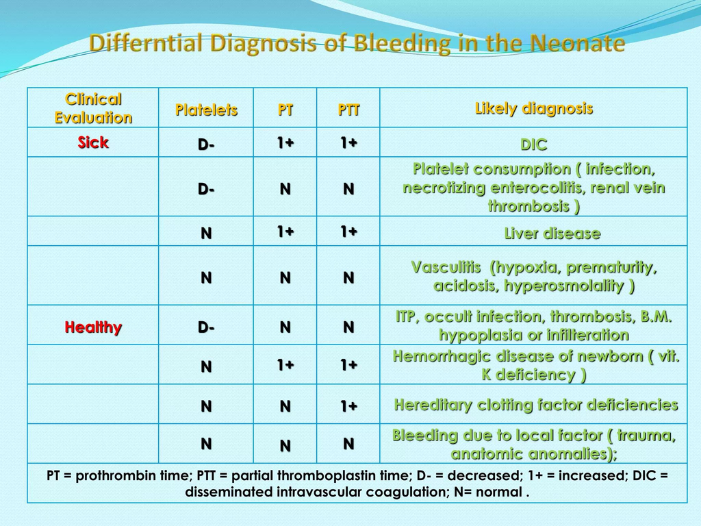
Prolonged PT/INR in neonates usually reflects abnormalities in the extrinsic and common coagulation pathways (Factors I, II, V, VII, X).
The causes can be grouped into physiologic, nutritional, hepatic, consumptive, inherited, and iatrogenic causes.


1. Physiological Causes

Physiologic immaturity of coagulation system

  • Normal neonates (especially preterms) have:
    • Lower vitamin K–dependent clotting factors
    • Reduced hepatic synthesis
  • Mild PT prolongation may occur in:
    • Prematurity
    • Very low birth weight infants

2. Vitamin K Deficiency (Most Important Cause)

Vitamin K Deficiency Bleeding (VKDB)

Most common clinically important cause of prolonged PT in neonates.

Why PT increases first?

Factor VII has the shortest half-life → PT prolongs earliest.

Risk factors

  • No vitamin K prophylaxis at birth
  • Exclusive breastfeeding
  • Maternal anticonvulsants:
    • Phenytoin
    • Phenobarbital
    • Carbamazepine
  • Maternal anti-TB drugs:
    • Rifampicin
    • Isoniazid
  • Malabsorption/cholestasis
  • Prolonged antibiotics

Types

TypeTiming
Early VKDB<24 hr
Classical VKDBDay 2–7
Late VKDB2 weeks–6 months

Labs

  • ↑ PT/INR (earliest finding)
  • Later ↑ aPTT
  • Normal platelets initially
  • Rapid correction after vitamin K

3. Liver Disease / Hepatic Dysfunction

Neonatal liver dysfunction reduces clotting factor synthesis.

Causes

  • Neonatal hepatitis
  • Biliary atresia
  • Cholestatic liver disease
  • Sepsis-associated liver dysfunction
  • Hypoxic ischemic injury
  • Metabolic liver disease
    • Galactosemia
    • Tyrosinemia

Labs

  • ↑ PT/INR
  • May not correct with vitamin K if severe hepatocellular failure

4. Disseminated Intravascular Coagulation (DIC)

Consumptive coagulopathy causing depletion of clotting factors.

Common neonatal triggers

  • Sepsis
  • Birth asphyxia
  • NEC
  • Shock
  • Severe hypoxia
  • Meconium aspiration
  • Severe hemolysis

Labs

  • ↑ PT
  • ↑ aPTT
  • ↓ fibrinogen
  • ↓ platelets
  • ↑ D-dimer/FDP

5. Sepsis

Can prolong PT due to:

  • Hepatic dysfunction
  • DIC
  • Cytokine-mediated coagulopathy
  • Vitamin K deficiency from poor feeding/antibiotics

6. Congenital Coagulation Factor Deficiencies

Factor VII deficiency

  • Isolated prolonged PT
  • Normal aPTT

Deficiencies affecting common pathway

  • Factor II deficiency
  • Factor V deficiency
  • Factor X deficiency
  • Fibrinogen disorders

Clues

  • Family history
  • Bleeding despite vitamin K
  • Persistent abnormal PT

7. Massive Bleeding / Dilutional Coagulopathy

Occurs after:

  • Massive transfusion
  • Exchange transfusion
  • Severe hemorrhage

Mechanism:

  • Dilution of clotting factors

8. Anticoagulant Exposure

Maternal drug exposure

  • Warfarin
  • Anticonvulsants
  • Anti-TB drugs

Neonatal medications

  • Heparin contamination of sample (usually affects aPTT more)

9. Cholestasis / Fat Malabsorption

Leads to poor vitamin K absorption.

Causes

  • Biliary atresia
  • Neonatal cholestasis
  • Cystic fibrosis
  • Intestinal disease

10. Laboratory / Sampling Errors

Always exclude before extensive workup.

Causes

  • Underfilled citrate tube
  • Clotted sample
  • Heparin contamination
  • Difficult sampling

Important High-Yield Pattern Recognition

PatternLikely Cause
Isolated ↑ PTVitamin K deficiency, Factor VII deficiency
↑ PT + ↑ aPTTLiver disease, DIC, severe VKDB
↑ PT + thrombocytopeniaDIC, sepsis
PT corrects after vitamin KVKDB
PT not correcting with vitamin KLiver failure or factor deficiency

High-Yield Neonatal Causes to Remember

  1. Vitamin K deficiency
  2. Sepsis
  3. DIC
  4. Liver disease/cholestasis
  5. Prematurity
  6. Congenital factor deficiency
  7. Maternal drug exposure

🚨 Increased PT/INR in Neonates — High Yield NICU Causes

🩸 Common causes of prolonged PT/INR in newborns:

▪️Vitamin K deficiency (most common)
▪️Sepsis
▪️DIC
▪️Liver disease / cholestasis
▪️Prematurity (physiologic immaturity)
▪️Congenital factor deficiencies (especially Factor VII)
▪️Maternal drugs
  • Anticonvulsants
  • Anti-TB drugs
  • Warfarin
▪️Massive bleeding / dilutional coagulopathy
▪️Laboratory sampling errors

⚡ High-yield tip:
➡️ PT rises EARLY in vitamin K deficiency because Factor VII has the shortest half-life.

🧠 Pattern to remember:
🔹 Isolated ↑ PT → Vitamin K deficiency / Factor VII deficiency
🔹 ↑ PT + ↑ aPTT → DIC or liver disease
🔹 Corrects after vitamin K → VKDB likely

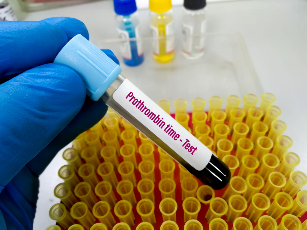
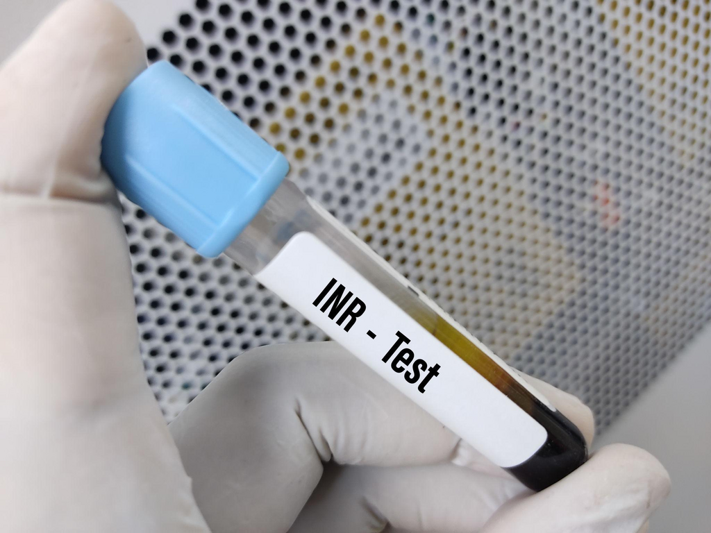
PT/INR evaluates the extrinsic coagulation pathway and is commonly used in neonatal coagulation assessment. (bmcpediatr.biomedcentral.com)

6 Well Known Step Pathogenesis of Apnea of Prematurity Based on Nelson and Clohery to Clear Your Concept

Apnea of prematurity (AOP) is a common condition where infants born before 37 weeks gestation experience breathing pauses lasting 15-20 seconds or more, often accompanied by slow heart rates (bradycardia) or low oxygen levels. It stems from an immature central nervous system and usually resolves on its own by 37–40 weeks postmenstrual age. Based on Cloherty and Stark’s Manual of Neonatal Care (9th Edition) and relevant pediatric algorithms and Nelson’s, here is a comprehensive summary of Apnea of Prematurity (AOP).

1. Definition and Classification

  • Definition: Apnea is the cessation of airflow. It is considered pathologic (an apneic spell) when:
    • Absence of airflow lasts 20 seconds or longer.
    • It is shorter than 20 seconds but accompanied by bradycardia (heart rate < 100 bpm) or hypoxemia (cyanosis or $SpO_2$ < 85–80%).
  • Classification:
    • Central: Total absence of inspiratory efforts (no diaphragmatic activity).
    • Obstructive: Inspiratory efforts persist, but airflow is blocked, usually at the pharyngeal level.
    • Mixed: A combination where airway obstruction precedes or follows central apnea. Most spells in preterm infants are mixed.

2. Incidence, Onset, and Duration

  • Incidence: Inversely related to gestational age (GA). It occurs in essentially all infants < 28 weeks’ GA and about 25% of those < 34 weeks’ GA.
  • Onset: Typically begins 1 to 2 days after birth. If spells do not occur within the first 7 days, AOP is unlikely to develop later unless triggered by other factors.
  • Duration: Usually ceases by 36 to 37 weeks’ postmenstrual age (PMA) in infants born at $\ge 28$ weeks, but frequently persists beyond term for those born more prematurely.

3. Pathogenesis (Underlying Mechanisms)

  • Developmental Immaturity: The primary cause is an immature central respiratory drive in the brainstem.
  • Sleep State: Spells are more frequent during active (REM) sleep, which is the predominant sleep state in preterms and is characterized by irregular breathing.
  • Chemoreceptor Response: Preterm infants have a decreased ventilatory response to increased $CO_2$ and may respond to hypoxia with hypoventilation rather than sustained hyperventilation.
  • Reflexes: Apnea can be triggered by stimulation of the posterior pharynx (e.g., vigorous suctioning), lung inflation, or fluid in the larynx.
  • Airway Mechanics: Poor muscle tone can lead to airway obstruction, especially during neck flexion or if there is nasal obstruction.
  • Note on GER: While gastroesophageal reflux is common in preterms, studies have not demonstrated an association between GER and AOP frequency.

4. Differential Diagnosis (Evaluation)

Apnea in a term infant or a “sick” preterm infant is always abnormal and requires looking for secondary causes:

  • Infection: Sepsis, meningitis, or necrotizing enterocolitis.
  • Metabolic Disorders: Hypoglycemia, hypocalcemia, or electrolyte imbalances (hyponatremia).
  • Neurologic: Intracranial hemorrhage (IVH), seizures, or birth asphyxia.
  • Impaired Oxygenation: PDA (Patent Ductus Arteriosus), anemia, or pneumonia.
  • Drugs: Maternal medications (magnesium, narcotics) or drug toxicity (e.g., phenobarbitone).

5. Management and Treatment

  • Monitoring: All infants < 35 weeks’ GA should be monitored for at least the first week. Monitor heart rate and $SpO_2$ in addition to respiration.
  • Immediate Action: Respond to the infant, not the monitor alarm. Most spells respond to tactile stimulation. If the infant is unresponsive, use bag-and-mask ventilation.
  • Positioning: Avoid extreme neck flexion or extension. Prone positioning may reduce apnea by stabilizing the chest wall.
  • Pharmacotherapy (Methylxanthines):
    • Caffeine Citrate: The drug of choice due to its long half-life (once-daily dosing), high therapeutic index, and lack of need for routine level monitoring.
    • Dosing: Loading dose of 20 mg/kg (10 mg/kg caffeine base), followed by a maintenance dose of 5–10 mg/kg daily.
    • Benefits: Reduces spells, the need for mechanical ventilation, and the risk of Bronchopulmonary Dysplasia (BPD).
  • Respiratory Support:
    • nCPAP (4–6 $cm H_2O$): Reduces mixed and obstructive spells by maintaining end-expiratory lung volume.
    • NIPPV: May be attempted if CPAP fails.
  • Other Considerations:
    • Blood Transfusion: May be considered if the hematocrit is < 25–30% and spells are frequent despite caffeine.
    • GER Treatment: Pharmacologic treatment of reflux (e.g., H2 blockers) is not recommended to treat AOP and may be harmful.

6. Discharge and Follow-up

  • Discharge Criteria: Infants should be free of significant apnea for 5 to 7 days after stopping caffeine.
  • Caffeine Offset: Because caffeine effects remain for up to a week, the “countdown” to discharge typically starts several days after the last dose.
  • Recurrence: Recurrent apnea can be triggered by viral illness, anesthesia, eye examinations, or immunizations. These infants should be monitored closely until at least 44 weeks’ PMA.
  • SIDS: A history of AOP does not increase the risk of Sudden Infant Death Syndrome (SIDS).

6 Important Things on Nursing care for an intubated infant and Child

Nursing care for an intubated infant or child requires a multi-system approach focused on airway security, meticulous monitoring, pulmonary hygiene, and developmentally supportive care to minimize complications and stress.

1. Airway Security and Tube Management

  • Tube Fixation and Position: The endotracheal tube (ETT) must be secured appropriately using waterproof tape or a specialized tube-securing device. The correct depth of insertion should be confirmed immediately via clinical assessment (equal bilateral air entry, symmetric chest rise) and subsequently by chest X-ray, which should show the tip between the T1 vertebra and the carina.
  • Positioning: Maintain the patient in a midline, neutral “sniffing” position with slight neck extension to prevent airway obstruction or accidental displacement. Infants and toddlers often require a towel roll beneath the shoulders to compensate for a large occiput and align the airway.
  • Emergency Preparedness (DOPE): Nurses must be vigilant for sudden deterioration using the DOPE mnemonic to identify causes: Displacement of the tube, Obstruction (e.g., by meconium or mucus), Pneumothorax, or Equipment failure. A manual ventilation system (T-piece resuscitator or self-inflating bag) and a spare ETT of the same size and one size smaller must be at the bedside at all times.

2. Pulmonary Hygiene

  • Judicious Suctioning: Routine suctioning is discouraged. It should be performed only when clinically indicated, such as visible/audible secretions, diminished chest wall movement (“wobble”), or worsening oxygenation/rising $CO_2$ suggesting an obstruction.
  • Suction Technique: Use in-line (closed) suctioning where possible to minimize circuit disruption, loss of lung volume (PEEP), and the risk of infection. Suction pressure should be maintained between 80 and 120 mmHg. Limit suctioning attempts to avoid inducing bradycardia or hypoxia.
  • Humidification and Warming: Inspired gases must be heated and humidified to prevent the drying of airways and secretions, which can lead to tube occlusion, and to avoid convective heat loss.

3. Monitoring and Assessment

  • Continuous Monitoring: Vital signs including heart rate (via ECG), respiratory rate, and oxygen saturation ($SpO_2$) must be monitored continuously. In neonates, an ECG is often more accurate than pulse oximetry for determining heart rate during periods of poor perfusion.
  • Ventilation Efficacy: Monitor for equal breath sounds and adequate, but not excessive, chest rise. Frequent blood gas analysis (arterial, capillary, or venous) is necessary to titrate ventilator settings. Non-invasive methods like transcutaneous $CO_2$ ($TcPCO_2$) monitoring or capnography may be used to identify trends.
  • Systemic Support: Monitor blood pressure, capillary refill time, and urine output (target 1.5–2 mL/kg/hr in infants) to ensure adequate perfusion while on positive pressure ventilation.

4. Developmental and Comfort Care

  • Minimal Handling: Coordinate care activities to allow for long periods of undisturbed rest, as frequent handling can lead to fluctuations in cerebral circulation and clinical deterioration.
  • Sedation and Analgesia: Provide adequate analgesia and sedation (e.g., morphine or fentanyl infusions) to prevent agitation, which can increase intracranial pressure and cause the patient to “fight” the ventilator. Neuromuscular blockade (paralysis) is rarely used but may be necessary for severe cases, such as meconium aspiration syndrome, to synchronize breathing.
  • Supportive Environment: Use “nesting” materials or swaddling to maintain a flexed, midline posture, which helps with self-regulation and reduces stress. Ensure a quiet environment by silencing alarms quickly and speaking in low tones.

5. Nutrition and Gastric Care

  • Gastric Decompression: An orogastric or nasogastric tube should be placed and left on “free drainage” to decompress the stomach. This prevents diaphragmatic splinting from swallowed air, which can compromise ventilation.
  • Enteral Feeding: Once the patient is hemodynamically stable, careful gastric tube feeding (preferably with breast milk) is not contraindicated and should be initiated as tolerated.

6. Weaning and Extubation Care

  • Assessing Readiness: Weaning should begin as soon as the primary disease process improves and the patient shows spontaneous breathing efforts. Standard indices for readiness include $FiO_2$ <50% and stable blood gases on minimal settings.
  • Extubation Support: Prior to extubation, provide suctioning and gentle chest physiotherapy. Provide humidified oxygen via nasal cannula or hood immediately following the procedure. In preterm infants, transitioning to nasal CPAP often helps prevent extubation failure.