
Table of Contents
Nursing care for an intubated infant or child requires a multi-system approach focused on airway security, meticulous monitoring, pulmonary hygiene, and developmentally supportive care to minimize complications and stress.
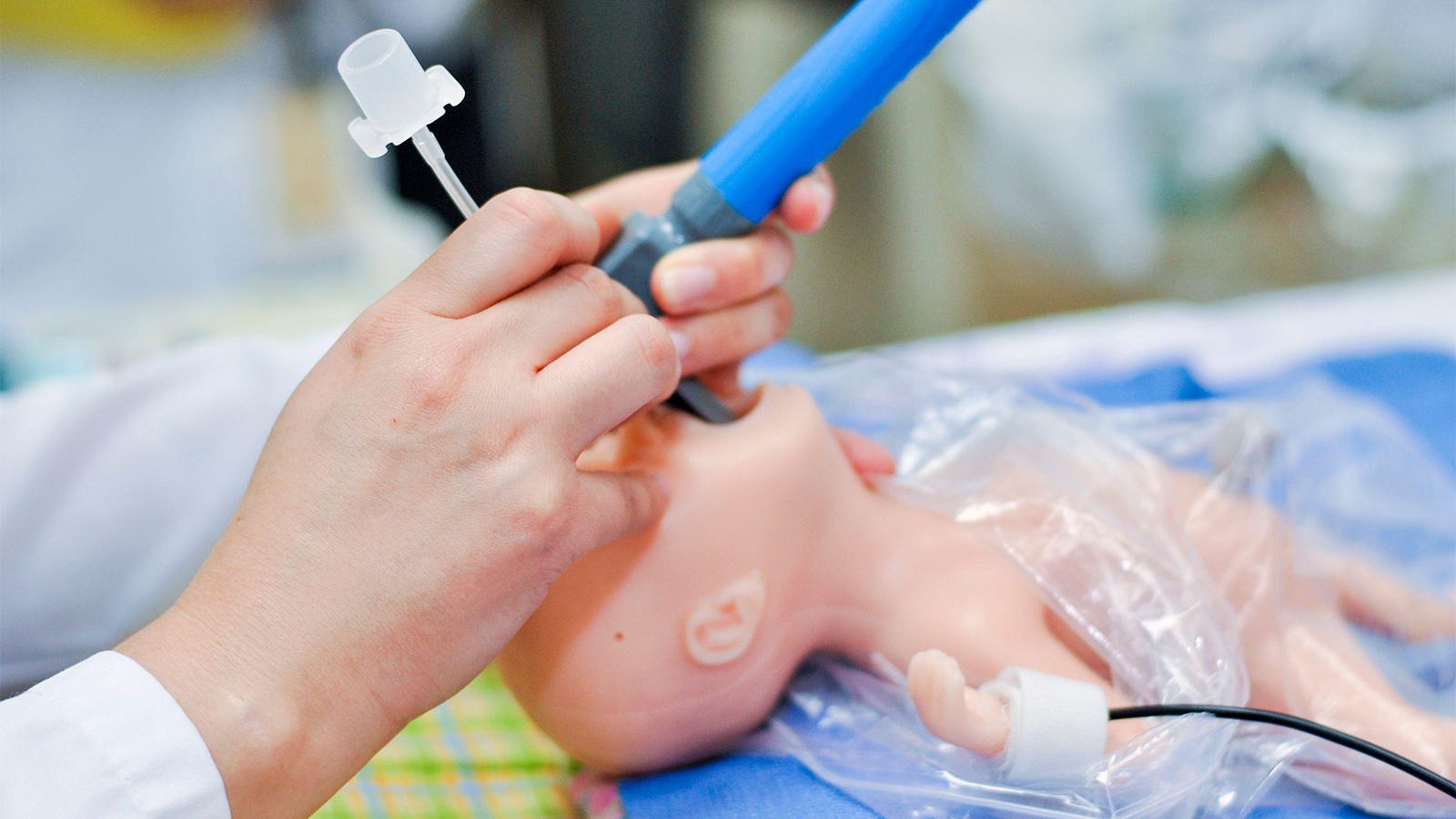
1. Airway Security and Tube Management
- Tube Fixation and Position: The endotracheal tube (ETT) must be secured appropriately using waterproof tape or a specialized tube-securing device. The correct depth of insertion should be confirmed immediately via clinical assessment (equal bilateral air entry, symmetric chest rise) and subsequently by chest X-ray, which should show the tip between the T1 vertebra and the carina.
- Positioning: Maintain the patient in a midline, neutral “sniffing” position with slight neck extension to prevent airway obstruction or accidental displacement. Infants and toddlers often require a towel roll beneath the shoulders to compensate for a large occiput and align the airway.
- Emergency Preparedness (DOPE): Nurses must be vigilant for sudden deterioration using the DOPE mnemonic to identify causes: Displacement of the tube, Obstruction (e.g., by meconium or mucus), Pneumothorax, or Equipment failure. A manual ventilation system (T-piece resuscitator or self-inflating bag) and a spare ETT of the same size and one size smaller must be at the bedside at all times.
2. Pulmonary Hygiene
- Judicious Suctioning: Routine suctioning is discouraged. It should be performed only when clinically indicated, such as visible/audible secretions, diminished chest wall movement (“wobble”), or worsening oxygenation/rising $CO_2$ suggesting an obstruction.
- Suction Technique: Use in-line (closed) suctioning where possible to minimize circuit disruption, loss of lung volume (PEEP), and the risk of infection. Suction pressure should be maintained between 80 and 120 mmHg. Limit suctioning attempts to avoid inducing bradycardia or hypoxia.
- Humidification and Warming: Inspired gases must be heated and humidified to prevent the drying of airways and secretions, which can lead to tube occlusion, and to avoid convective heat loss.
3. Monitoring and Assessment
- Continuous Monitoring: Vital signs including heart rate (via ECG), respiratory rate, and oxygen saturation ($SpO_2$) must be monitored continuously. In neonates, an ECG is often more accurate than pulse oximetry for determining heart rate during periods of poor perfusion.
- Ventilation Efficacy: Monitor for equal breath sounds and adequate, but not excessive, chest rise. Frequent blood gas analysis (arterial, capillary, or venous) is necessary to titrate ventilator settings. Non-invasive methods like transcutaneous $CO_2$ ($TcPCO_2$) monitoring or capnography may be used to identify trends.
- Systemic Support: Monitor blood pressure, capillary refill time, and urine output (target 1.5–2 mL/kg/hr in infants) to ensure adequate perfusion while on positive pressure ventilation.
4. Developmental and Comfort Care
- Minimal Handling: Coordinate care activities to allow for long periods of undisturbed rest, as frequent handling can lead to fluctuations in cerebral circulation and clinical deterioration.
- Sedation and Analgesia: Provide adequate analgesia and sedation (e.g., morphine or fentanyl infusions) to prevent agitation, which can increase intracranial pressure and cause the patient to “fight” the ventilator. Neuromuscular blockade (paralysis) is rarely used but may be necessary for severe cases, such as meconium aspiration syndrome, to synchronize breathing.
- Supportive Environment: Use “nesting” materials or swaddling to maintain a flexed, midline posture, which helps with self-regulation and reduces stress. Ensure a quiet environment by silencing alarms quickly and speaking in low tones.
5. Nutrition and Gastric Care
- Gastric Decompression: An orogastric or nasogastric tube should be placed and left on “free drainage” to decompress the stomach. This prevents diaphragmatic splinting from swallowed air, which can compromise ventilation.
- Enteral Feeding: Once the patient is hemodynamically stable, careful gastric tube feeding (preferably with breast milk) is not contraindicated and should be initiated as tolerated.
6. Weaning and Extubation Care
- Assessing Readiness: Weaning should begin as soon as the primary disease process improves and the patient shows spontaneous breathing efforts. Standard indices for readiness include $FiO_2$ <50% and stable blood gases on minimal settings.
- Extubation Support: Prior to extubation, provide suctioning and gentle chest physiotherapy. Provide humidified oxygen via nasal cannula or hood immediately following the procedure. In preterm infants, transitioning to nasal CPAP often helps prevent extubation failure.