Daily Archives: November 1, 2025

uti

Complicated Vs Uncomplicated UTI (Notes) – Clinical Practice and Diagnosis


1. Definition

TypeDefinition
Uncomplicated UTI
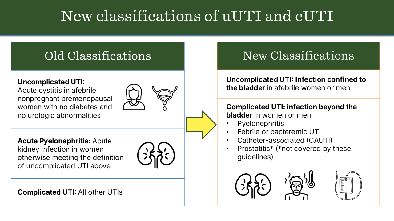
Infection of the lower urinary tract (bladder and urethra) occurring in an otherwise healthy, non-pregnant woman with a structurally and functionally normal urinary tract.
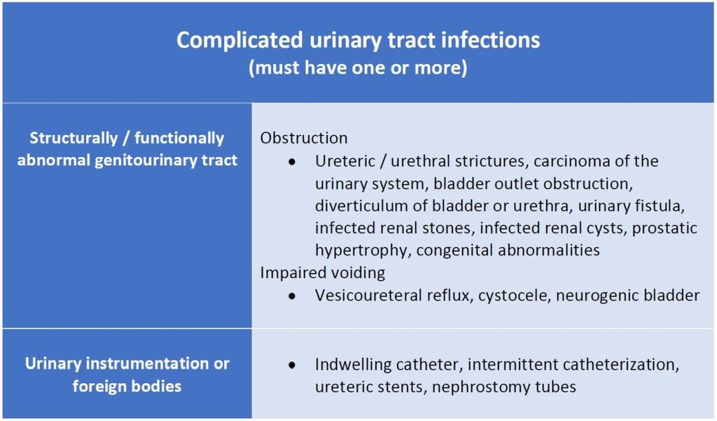
Complicated UTIUTI occurring in individuals with structural or functional abnormalities of the urinary tract, comorbidities, or other risk factors that increase risk of treatment failure or recurrence.

2. Typical Patients

TypeCommon in
UncomplicatedHealthy, premenopausal, non-pregnant females
ComplicatedMales, pregnant females, children, elderly, diabetics, catheterized patients, immunocompromised patients

3. Etiology

TypeCommon PathogensSpecial Pathogens
UncomplicatedE. coli (≈80–90%), Staphylococcus saprophyticusRarely Klebsiella, Proteus
ComplicatedE. coli, Klebsiella, Proteus, Enterobacter, Pseudomonas, Enterococcus, CandidaOften multidrug-resistant (MDR) organisms

4. Predisposing / Risk Factors

Uncomplicated:

  • Female gender (short urethra)
  • Sexual activity
  • Spermicides

Complicated:

  • Structural: Stones, strictures, obstruction (BPH, vesicoureteral reflux)
  • Functional: Neurogenic bladder, indwelling catheter
  • Systemic: Diabetes mellitus, immunosuppression
  • Male gender
  • Pregnancy

5. Clinical Presentation

TypeFeatures
UncomplicatedDysuria, frequency, urgency, suprapubic pain, no systemic signs
ComplicatedMay have fever, chills, flank pain, systemic toxicity, sepsis, poor response to therapy

6. Investigations

TypeLab Approach
UncomplicatedUrinalysis ± urine culture (often empirical treatment)
ComplicatedUrine culture mandatory, imaging (USG, CT KUB) if obstruction suspected, blood cultures if febrile

7. Treatment

TypeApproach
UncomplicatedShort-course oral antibiotics (3–5 days): Nitrofurantoin, TMP-SMX, Fosfomycin, Pivmecillinam
ComplicatedLonger course (7–14 days), guided by culture; IV therapy if severe (Ceftriaxone, Piperacillin-tazobactam, Carbapenems for MDR); treat underlying cause (remove catheter, relieve obstruction)

8. Prognosis

TypePrognosis
UncomplicatedExcellent, low recurrence with proper hygiene
ComplicatedRisk of recurrence, sepsis, renal scarring, abscess

9. Example Cases

ScenarioType
25-year-old woman with dysuria, no comorbiditiesUncomplicated
60-year-old diabetic man with fever, flank painComplicated
Pregnant woman with bacteriuriaComplicated
Patient with indwelling Foley catheter and feverComplicated

Summary Table

FeatureUncomplicatedComplicated
HostHealthy femaleAny comorbidity or abnormality
SiteLower UTI (cystitis)Any (cystitis, pyelonephritis, sepsis)
OrganismsUsually E. coliPolymicrobial, resistant organisms
TherapyShort courseLong course, guided by culture
PrognosisExcellentVariable, risk of recurrence

Aphthous Ulcers: Step-by-Step Workup and Management

1. Clinical Assessment

History

  • Onset, duration, frequency, and number of ulcers
  • Pain severity, triggers (trauma, stress, certain foods)
  • Systemic symptoms: fever, diarrhea, joint pain, fatigue
  • Family history of similar lesions
  • Medications (NSAIDs, beta-blockers)
  • Previous treatments and response

Examination

  • Location: non-keratinized mucosa (buccal, labial, ventral tongue, floor of mouth)
  • Size:
    • Minor (<1 cm) – most common
    • Major (>1 cm, deeper, may scar)
    • Herpetiform (multiple small ulcers)
  • Number: single or multiple
  • Appearance: round/oval, yellow-white pseudomembrane with erythematous halo
  • Rule out: secondary causes (herpes simplex, trauma, systemic disease)

2. Investigations (If atypical or recurrent)

  • Basic labs: CBC, iron studies, vitamin B12, folate (nutritional deficiencies)
  • CRP/ESR: if systemic symptoms
  • Serology: celiac disease (tTG-IgA), HIV (if risk factors)
  • Biopsy: rare, only if persistent >3 weeks, atypical, or suspected malignancy
  • Microbiology: rarely needed; ulcers are typically non-infectious

3. Classification

TypeFeaturesManagement Focus
Minor<1 cm, 1–5 lesions, heal 7–14 daysSymptomatic relief
Major>1 cm, deep, persistent, may scarSystemic therapy possible
HerpetiformMultiple, small, coalescing, painfulCombination topical/systemic therapy

4. Management

A. General Measures

  • Avoid triggers: spicy/acidic foods, trauma
  • Good oral hygiene
  • Avoid sodium lauryl sulfate in toothpaste
  • Stress management
  • Nutritional supplementation if deficient

B. Topical Therapy (First-line for minor ulcers)

  • Topical corticosteroids:
    • Triamcinolone acetonide 0.1% in orabase
    • Fluocinonide 0.05% gel
  • Topical anesthetics:
    • Lidocaine 2% gel for pain relief
  • Protective agents:
    • Orabase, hydroxypropyl cellulose films

C. Systemic Therapy (For severe, recurrent, or major ulcers)

  • Oral corticosteroids:
    • Prednisone short course (e.g., 30–60 mg/day tapering)
  • Immunomodulators (refractory cases):
    • Colchicine, thalidomide, dapsone
  • Address underlying systemic disease:
    • Nutritional deficiencies, IBD, Behçet’s disease, etc.

D. Adjunctive Therapy

  • Antimicrobial mouth rinses: chlorhexidine gluconate
  • Pain control: NSAIDs (if not contraindicated)
  • Laser therapy (experimental for pain reduction and healing)

5. Follow-Up

  • Minor ulcers: usually self-limiting; review if >3 weeks
  • Recurrent or major ulcers: monitor frequency, severity, and response to therapy
  • Evaluate for systemic disease if persistent or atypical

Approach to a Child with Wheeze

1. Definition

  • Wheeze is a high-pitched, musical, continuous sound produced by airflow through narrowed airways, typically during expiration.
  • Indicates airway obstruction at any level (from trachea to small bronchi).

2. Initial Assessment

A. ABC Assessment

  • Airway: Ensure patency; look for obstruction (secretions, foreign body).
  • Breathing:
    • Respiratory rate, effort (retractions, nasal flaring, grunting)
    • Oxygen saturation (SpO₂)
    • Presence and distribution of wheeze
    • Work of breathing (mild / moderate / severe)
  • Circulation: Heart rate, capillary refill time, BP, color.

B. Severity Assessment

SeverityFeatures
MildSpeaking in sentences, SpO₂ > 94%, mild wheeze
ModerateBreathless, feeding difficulty, SpO₂ 90–94%
SevereCannot talk/feed, SpO₂ < 90%, exhaustion, cyanosis, silent chest

3. Focused History

AspectKey Points
Onset & CourseSudden (foreign body) vs gradual (infection/asthma)
Frequency/PatternRecurrent vs first episode
TriggersViral infection, allergen, exercise, cold air, smoke
Associated SymptomsFever, cough, coryza, vomiting, feeding difficulty
Past Medical HistoryPrevious wheezing, atopy, eczema, prematurity, GERD
Family/Social HistoryAsthma, allergies, smoking, housing
Drug historyRecent medications, response to bronchodilators

4. Physical Examination

  • General: Distress, cyanosis, growth, hydration.
  • Respiratory system:
    • Chest expansion, use of accessory muscles
    • Percussion (hyperresonant / dull)
    • Auscultation:
      • Wheeze: polyphonic (asthma/viral), monophonic (focal obstruction)
      • Air entry: symmetrical or reduced
      • Crackles: suggest infection or bronchiolitis
  • Cardiac: Murmurs (congenital lesions)
  • Other systems: Skin (eczema), ENT (allergic rhinitis), clubbing.

5. Differential Diagnosis

Age GroupCommon Causes
Infant (<1 yr)Bronchiolitis, congenital airway anomalies, aspiration, GERD
Toddler (1–5 yr)Viral-induced wheeze, foreign body aspiration, asthma
Older child (>5 yr)Asthma, allergic bronchitis, chronic suppurative lung disease

6. Investigations

(Usually guided by clinical picture; many cases diagnosed clinically)

  • Pulse oximetry – essential.
  • Chest X-ray – if first episode, focal findings, suspicion of foreign body/pneumonia.
  • Blood tests – if severe or infection suspected.
  • Allergy testing / IgE – in recurrent or atopic cases.
  • Spirometry / Peak Flow – for older cooperative children (asthma diagnosis).
  • Bronchoscopy – if persistent localized wheeze or suspicion of foreign body.

7. Management

A. Immediate Management

  • Mild/Moderate:
    • Nebulized or inhaled salbutamol (via spacer or nebulizer)
    • Oxygen if SpO₂ < 94%
    • Oral prednisolone if known asthma or recurrent wheeze
  • Severe / Life-threatening:
    • High-flow oxygen
    • Nebulized salbutamol ± ipratropium bromide
    • IV steroids (hydrocortisone)
    • IV magnesium sulfate / aminophylline / salbutamol if poor response
    • Consider PICU referral

B. Underlying Cause

  • Bronchiolitis – supportive (O₂, fluids, suction)
  • Foreign body – urgent ENT/pulmonary referral
  • Asthma – follow stepwise management (as per BTS/SIGN or GINA)

8. Long-term Management

  • Identify triggers and educate parents on avoidance.
  • Asthma education: inhaler technique, action plan.
  • Follow-up to reassess control and adjust therapy.

9. Red Flags

  • Silent chest, exhaustion, cyanosis
  • Poor air entry or asymmetry
  • Persistent localized wheeze
  • Failure to thrive or recurrent pneumonia
  • Sudden onset without infection (→ foreign body)

10. Summary Table

StepKey Action
1ABC and severity assessment
2Focused history
3Physical examination
4Consider differential diagnoses
5Targeted investigations
6Manage acutely + treat cause
7Educate and follow-up