Table of Contents
For Japanese Encephalitis (JE), CT findings are not always very sensitive early on, but there are classical (almost pathognomonic) patterns you should remember:
Key Pathognomonic CT Finding
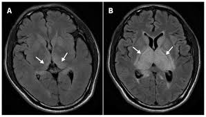
👉 Bilateral thalamic hypodensities


- Most characteristic feature
- Typically bilateral, symmetrical low-density lesions
- May show hemorrhagic changes in severe cases
Other Supporting CT Findings
(Not strictly pathognomonic but highly suggestive in right clinical setting)
- Basal ganglia involvement
- Midbrain involvement
- Brainstem lesions
- Cerebellar involvement (less common)
- Diffuse cerebral edema in severe cases
Important Clinical Correlation
- JE has a predilection for deep gray matter, especially thalamus
- Similar pattern can be seen in:
- West Nile encephalitis
- Acute necrotizing encephalopathy
👉 So diagnosis = CT pattern + epidemiology + CSF + serology (IgM)
Exam Pearl (VERY HIGH-YIELD)
“Bilateral thalamic lesions on CT/MRI = Think Japanese Encephalitis first (especially in endemic areas like Nepal/India)”